
Figures & data
Table I Incubation conditions and time required to induce sporulation in Phialocephala spp.
Fig. 1 Bayesian 50% majority rule ITS and RPB1 consensus trees containing representative Mollisia and Phialocephala isolates: culture collection, GenBank or JB Tanney personal collection accession numbers are followed by the species name (T = ex-type; E = ex-epitype). All branches have Bayesian posterior probability values of 1.0; values lower than 1.0 are presented at nodes. The trees were rooted to Loramyces macrosporus and bars indicate expected changes per site per branch.
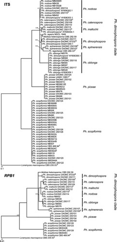
Fig. 2 Colony morphologies of representative isolates, 2 wk after inoculation on MEA at 20 C in the dark (T = ex-type, E = ex-epitype).
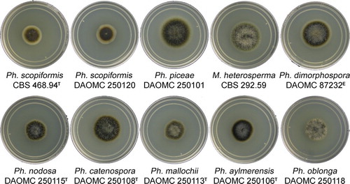
Fig. 3 A. Mature Phialocephala catenospora apothecia on 4 mo old culture (DAOMC 250108) on MEA. B–D. Synnemata of Ph. oblonga. E–G. Ph. dimorphospora conidiophores. H–N. Ph. catenospora synanamorphs. O–S. Ph. nodosa microsclerotia. T–X. Ph. aylmerensis conidiophores. Bars = 10 μm.
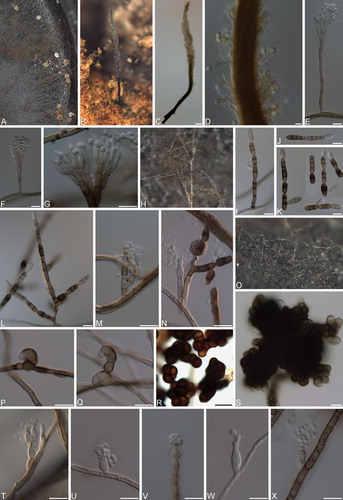
Fig. 4 Phialocephala scopiformis. A–C. Apothecia on fallen decaying Picea rubens branches; D. Vertical section of apothecium. E–F. Marginal cells. G. Vertical section displaying ectal and medullary excipulum. H–I. Paraphyses displaying refractive vacuole bodies. J. Asci with hemiamyloid tips in Lugol’s solution after KOH pretreatment. K. Discharged and germinating ascospores. Bars: D = 500 μm, E = 20 μm, F–K = 10 μm.
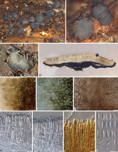
Fig. 5 Phialocephala piceae. A–C. Apothecia erumpent on fallen decaying Acer saccharum branches. D. Vertical section of apothecium. E–F. Marginal cells. G. Vertical section showing ectal and medullary excipulum. H. Ectal excipulum cells. I–J. Paraphyses displaying refractive vacuole bodies. K. Immature asci. L. Ascus with hemiamyloid tip in Lugol’s solution after KOH pretreatment. M. Ascospores. Bars: D = 500 μm, E–M = 10 μm.
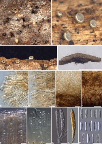
Fig. 6 Phialocephala nodosa. A–D. Apothecia on fallen decaying Acer saccharum branches. E. Vertical section showing margin and paraphyses with refractive vacuole bodies. F. Ectal excipulum. G–H. Marginal cells. I. Ectal excipulum cells. J. Subicular hyphae with thick gelatinous sheath. K. Paraphyses with refractive vacuole bodies. L–M. Asci with hemiamyloid tips in Lugol’s solution after KOH pretreatment. N. Ascospores under phase contrast. O. Ascospores under DIC. Bars = 10 μm.
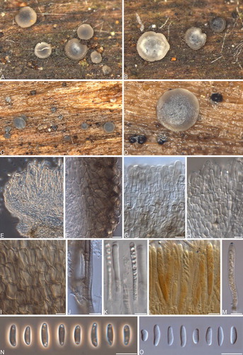
Fig. 7 Phialocephala catenospora. A–C. Apothecia on fallen decaying Betula papyrifera branch. D. Vertical section of apothecium. E. Vertical section of apothecial margin. F, G. Ectal and medullary excipulum. H. Ectal excipulum cells. I. Vertical section of apothecium with subicular hyphae in host tissue. J. Refractive vacuole bodies of paraphyses under DIC. K. Refractive vacuole bodies of paraphyses under phase contrast. L. Ectal excipulum cells mounted in H2O. M. Ectal excipulum cells in KOH. N. Asci. O–P. Asci with hemiamyloid tips in Lugol’s solution after KOH pretreatment. Q. Ascospores. Bars = 10 μm.
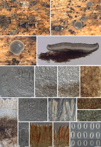
Fig. 8 Phialocephala mallochii. A–D. Apothecia on decaying Betula alleghaniensis log. E, F. Vertical sections of apothecia. G. Marginal cells. H. Ectal excipulum cells. I. Marginal cells with refractive vacuole bodies. J. Ectal and medullary excipulum. K. Refractive vacuole bodies of paraphyses. M. Ectal and medullary excipulum. N. Refractive vacuole bodies of paraphyses. O. Ascus. P. Ascus with hemiamyloid tip in Lugol’s solution after KOH pretreatment. Q. Ascospores. Bars: E = 500 μm, F–Q = 10 μm.
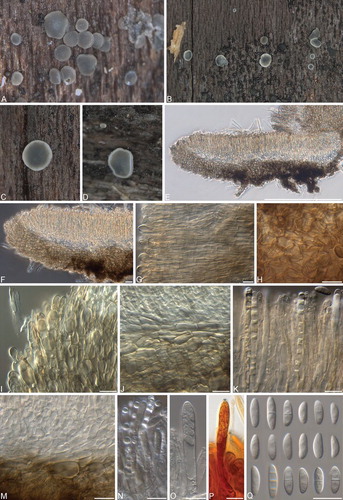
Fig. 9 Phialocephala oblonga. A–D. Apothecia on decayed Acer saccharum log. E, F. Vertical sections of apothecia. G. Vertical section of apothecial margin. H. Marginal cells with refractive vacuole bodies. I. Vertical section showing subicular hyphae in host tissue. J. Asci. K, L. Asci and paraphyses with refractive vacuole bodies. M, N. Asci with hemiamyloid tips in Lugol’s solution after KOH pretreatment. O. Ascospores under DIC (top three rows) and light microscopy (bottom row). Bars: E, F = 500 μm; G–O = 10 μm.
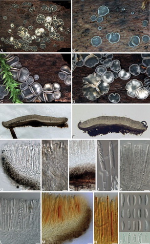
Fig. 10 Phialocephala aylmerensis. A–D. Apothecia on decaying Betula papyrifera wood. E, F. Vertical sections of apothecia. G. Subicular hyphae. H, I. Ectal excipulum cells near base (H) and toward flanks (I). J, K Marginal cells. L. Asci and paraphyses. M. Mature ascus containing ascospores. N. Asci with hemiamyloid tips in Lugol’s solution after KOH pretreatment. O. Ascospores under DIC. P. Ascospores under phase contrast. Bars: E = 500 μm, F = 100 μm, G–L, N = 10 μm, M, O, P = 5 μm.
