928
Views
0
CrossRef citations to date
0
Altmetric
Paper

Histological alterations of intestinal villi and epithelial cells after feeding dietary sugar cane extract in piglets

, , , , , , , & show all
Article: e43 | Received 06 Oct 2010, Accepted 19 Apr 2012, Published online: 18 Feb 2016

Figures & data

Table 1 Feed ingredients and chemical composition of basal commercial pre-starter and starter mush diets of piglets.
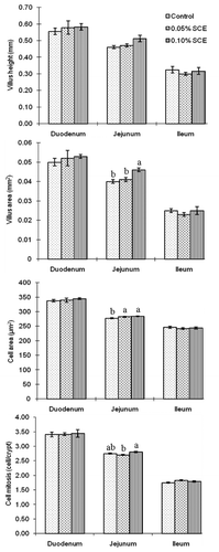
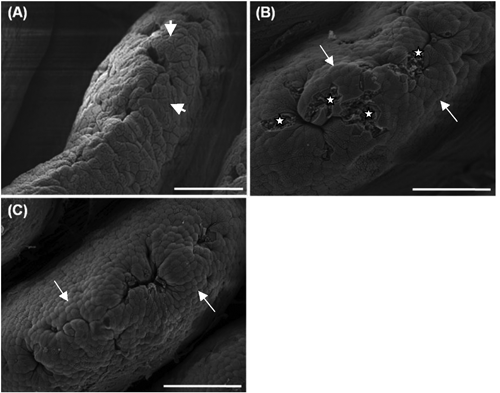
Figure 1 Intestinal villus height, villus area, cell area and cell mitosis of the duodenum, jejunum and ileum in piglets fed a basal diet with 0% (control), 0.05% and 0.10% dietary sugar cane extract (SCE).
Figure 2 Duodenal villus apical surface of piglets fed a basal diet with 0% (control; A), 0.05% (B), and 0.10% (C) dietary sugar cane extract (SCE). Scale bar: 50 µm. Control has faintly protuberated cells (short arrows), but SCE groups have clearly protuberated cells (long arrows). In 0.05 % SCE group, deeper cells at the sites of recently exfoliated cells (stars) appear.
Figure 3 Jejunal villus apical surface of piglets fed a basal diet with 0% (control; A), 0.05% (B), and 0.10% (C) dietary sugar cane extract (SCE). Scale bar: 50 µm. Much more developed cell clusters (arrow heads) can be seen in 0.05% group that in 0.10% group.
Figure 4 Ileal villus apical surface of piglets fed a basal diet with 0% (control; A), 0.05% (B), and 0.10% (C) dietary sugar cane extract (SCE). Scale bar: 50 µm.