715
Views
0
CrossRef citations to date
0
Altmetric
Paper

Neuroglial Cells in Long-Term Primary Cultures from the Gilthead Seabream (Sparus Aurata L.): New Functional In Vitro Model from Bony Fish Brain

, , , &
Article: e5 | Received 27 Aug 2012, Accepted 14 Nov 2012, Published online: 18 Feb 2016

Figures & data

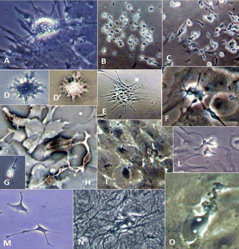
Figure 1. Morphological characterisation of SaGliPs cultures. Phase contrast microscopy images showing development and differentiation of Sparus aurata primary neuroglial cells (SaGliPs) in culture. (A) Morphology of radial glia progenitors (RGPs) showing a cluster of newly synthesised neural cell progenitors; 4-day old culture (400x). (B) Morphology of freshly isolated staminal neural cells (SNc) with the characteristic ovoid shape; 1-day old culture (200x). (C) Early elongated attached SNc after 2 days (200x). (D and D’) Highly branched fibrous astrocytes (200x). (E) RD colony at Day 6 of culture during initial process elongation (200x). (F) Morphology of neuron precursor cells (NPC) which generate neurons but not glia; colony showing a cluster of newly synthetized neurons (200x). (G) Early elongated attached neuron progenitor after 2 days (200x). (H) Negative inverted phase contrast image showing newly formed neurons from NPC in 6-day old culture (200x). (I) Astrocyte precursors, arrow indicates the emergence site of filopodia protrusion; 2-week old culture (200x). (L) First growing cluster of RD at Day 3 of culture (200x). (M) Formation of connections between two branched oligodendrocyte precursors (OPCs) at early stages (400x). (N) Morphology of microglial cells highly branched over a monolayer of astrocytes, 4-week old culture (200x). (O) Newlyformed RD cell resembling tanycyte; 2-week old culture (200x).
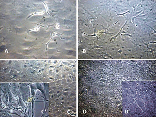
Figure 2. Characterisation of astrocyte monolayer. Phase contrast microscopy images showing the monolayer development and differentiation of primary neuroglial cell cultures isolated from the brain of Sparus aurata (SaGliPs). (A) Neural elongated stem cells (NS) at Day 4 (black arrows) and (B) while generating neurons (yellow arrows) and astrocytes (stellate cells) in 1-week old cultures. (C) Primary neuroglial cultures in enriched medium firstly differentiated in mature astrocyte cells forming a monolayer, and (C‘) generating neurons after 2 weeks of culture. (D) Monolayer of astrocyte cells with dense dendrite and neurite branching, and neuron bodies (D’) after 4 weeks of culture (200x).
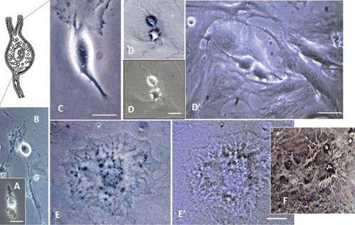
Figure 3. Oligodendrocyte lineage cell development and differentiation timing in neuroglial primary cultures from the brain of Sparus aurata (SaGliPs). Phase contrast microscopy images showing the lineage development of olygodendrocytes. (A) Neural stem cell (NS); 1-day old cultures. (B) Glial-restricted precursor (GRP) cells GRP; 2-day old cultures. (C) Prooligodendrocyte precursor (pOPC); 3-day old cultures. (D and D’) Oligodendrocyte precursors (OPCs); 1-week old cultures. (E and E’) Mature Oligodendrocyte (mOL); 3-week old cultures. mOL showed a round cell body enriched of cytoplasmic granules (E and E’), and a dense filamentous meshwork in the peripheral areas often enwrapping other neuroglial processes (F). A-D’, scale bar: 20 μ m; E-E’, scale bar: 50 μ m.
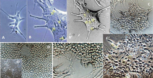
Figure 4. Radial glia development and differentiation from primary neuroglial cultures from the brain of Sparus aurata (SaGliPs). Phase contrast microscopy images showing radial glia progeny from neural stem cells. (A) Morphology of radial glia staminal progenitor; emergence sites of first protrusion end (black arrow) (200x). (B and B’) Enlarged detail of multipotent colony of radial glia; early stages at the site of emergence of growth cones of radial processes (yellow arrows) (400x). (C) Phase contrast image presented as inverse phase micrographs of panel C’ original image showing a rosette-like colony of radial glia after 2 weeks of culture (200x). (C’) An enlargement of some cells (C) showing detail of the radial colony with areas of active neuronal proliferation (asterisks), and first dividing cells generating neurons (yellow arrows) (400x). (D and E) Long radial glial fascicles of fibres running at the midline of the monolayer attempting to fill empty spaces (200x).
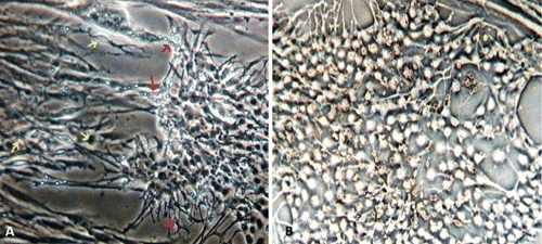
Figure 5. Proliferation of neurons in primary neuroglial cell cultures from the brain of Sparus aurata (SaGliPs). Phase contrast microscopy images showing the neural cell propagating in enriched differentiation medium supplemented with EGF, insulin, L-glutamine, hydrocortisone. (A) Phase contrast microscopy pictures showing radial glia in 2-week old cultures highly enriched in neurons (red arrows) dividing from neural stem cells; pro-oligodendrocytes precursor (yellow arrows) (200x). (B) Negative inverted image of enlarged area of the central zone of radial glia colony showing an intense granular production of neurons (200x).
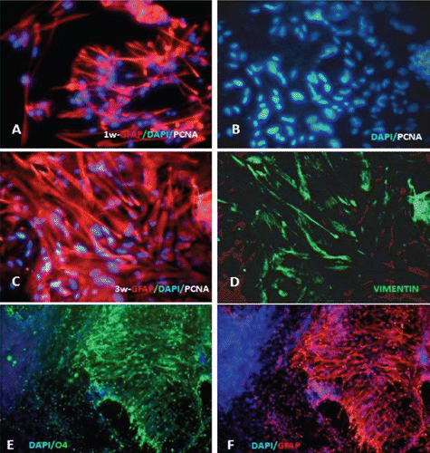
Figure 6. Indirect and direct immunofluorescence staining focusing astrocyte growth and differentiation in SaGliPs. (A) Intense fluorescence for GFAP at the tips of the astrocytes processes after 1 week of culture (x200). (B) DAPI counterstained nuclei corresponding to the same image positively labelled with GFAP and vimentin (C and D) (B-D, 2-week old cultures; 400x). The proliferative capacity of cells was shown by mitotic nuclei (white/cyan) doublestained with Ab anti-PCNA-FITC and counterstained with DAPI (blue). Radial glia stained immunopositive for both glial fibrillary acidic protein (GFAP) (C) and vimentin (D); 3-week old cultures. The DAPI and TRITC-fluorescence filter channels were acquired as digital images separately. The superimposition of two channels DAPI/TRITC was seen as merge. (E) Labelling with anti-O4 for oligodendrocytes (green) and DAPI (blue) to label cell nuclei. (F) Astrocytes immunostained with GFAP (red), and DAPI (blue) to label cell nuclei. (E and F) Co-localised areas of oligodendrocytes and astrocytes near enveloping axon fibres (200x).
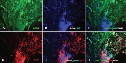
Figure 7. Double immunofluorescence demonstrating co-localisation of neurons and astrocytes within the radial glial colony. (A-C) Two-month old cultures showed actively replicating neurons (UCHL1+/DAPI+/PCNA+) inside the centre of the colony of radial glia (GFAP+). (D-F) The outer boundary of the radial glia colony showed (D) differentiated astrocytes with long GFAP+ filopodia and neuronal fibres (A, C and F), whereas in the inner zone there are clones of mitotic radial glia (PCNA+/DAPI+/GFAP+). (F) Several clusters of proliferative cells were observed; in particular, the central zone showed an intense granular production of neurons (PCNA+/UCHL1+). Radial glia remained mitotically active throughout the 2 months of culture. Original magnification: 200x.
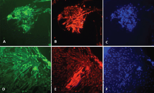
Figure 8. Radial glial progenitors can generate neurons and astrocytes in vitro. Long-term cultures of SaGliPs during neuronal differentiation efficiently generate neurons (A), from the colony of radial glia, immunostained with UCHL1 (highly specific to neurons) and astrocytes (B) that exhibit GFAP immunoreactivity. (C) Nuclei show the blue DAPI counterstaining. (AC) 2-week old cultures; (D-F) 2-month old cultures; (A-F) labelled with previous antibody (200x).
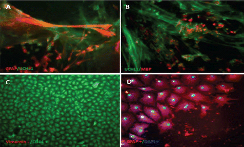
Figure 9. Double immunofluorescence demonstrating co-localisation of astrocytes and oligodendrocytes grown onto axons. Immunofluorescence staining focusing GFAP positive astrocytes differentiated into large cells with many filopodia while contacting multiple axons in order to sustain oligodendrocytes during the myelination process. Pictures in immunofluorescence staining as marked. (A) Glial fibrillary acidic protein (GFAP), the intermediate filament protein highly expressed in astrocytes (red), and UCHL1 (green) in seabream neuroglial cultures (200x). (B) Oligodendrocyte progenitor cells (OPC) visualised on neuron fibres. UCHL1 antibody was used to label neurons. Myelinating oligodendrocytes (labeled red) were immunostained with anti-myelin basic protein (MBP) (400x). (C) Monolayer of vimentinnegative astrocytes immunoreactive for CD44 marker of astrocyte lineage at 1 month (200x). (D) GFAP-immunoreactive mature astrocytes after 1 month of culture with the typical stellate shape and counterstained nuclei DAPI (400x).
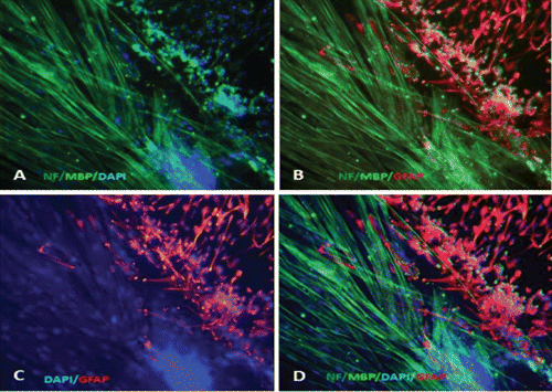
Figure 10. Immunocytochemistry analysis showing SaGliPs 3-week old cultures stained for several neuroglial specific markers. (A) Cells immunostained for labelling neurofilaments (NF) (visualised in axons, green), with DAPI (blue) to label cell nuclei, and anti MBP for myelinating oligodendrocytes. (B) Astrocytes were immunostained with GFAP (red), NF to label axons, and MBP to label OPCs. (C) Astrocytes immunostained with GFAP (red), and DAPI (blue) to label cell nuclei. (D) Merged photos displaying co-localisation of MBP and GFAP near NF positive axons. Myelinating oligodendrocytes co-localise with astrocyte while wrapping axons. Original magnification: 200x.