459
Views
7
CrossRef citations to date
0
Altmetric
Paper

Immunocytochemical Localisation of Phospholipid Hydroperoxide Glutathione Peroxidase in Bull’s Spermatogenic Cells

, , , &
Article: 3483 | Received 29 May 2014, Accepted 28 Jul 2014, Published online: 17 Feb 2016

Figures & data

Figure 1. Immunostaining of phospholipid hydroperoxide glutathione peroxidase in bull testis. Strongly stained cytoplasm of the maturing sperm cells localised in the apical compartment of the seminiferous tubules. Bar=20 μm.
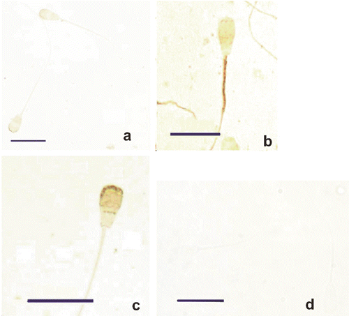
Figure 2. Immunostaining of phospholipid hydroperoxide glutathione peroxidase in ejaculated spermatozoa. Weak staining localised in the head and mid-piece portion (a). Immunostaining of the sperm mid-piece (b) and sperm head (c) increased by treatment with 6M guanidine-HCl and 10 mM glutathione, respectively. Positivity missing in control sections (d). Bars=10 μ.
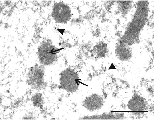
Figure 3. Immunoelectron microscopic localisation of phospholipid hydroperoxide glutathione peroxidase in spermatogenic cells of bull testis: spermatogonia. Gold particles showing phospholipid hydroperoxide glutathione peroxidase antigenic sites are present in mitochondria (arrows) and cytoplasmic matrix (arrow-heads). Bar=0.5 μ.
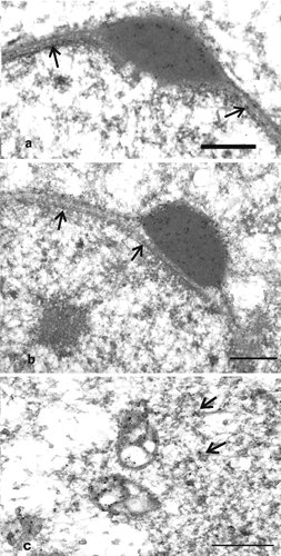
Figure 4. Immunoelectron microscopic localisation of phospholipid hydroperoxide glutathione peroxidase in spermatogenic cells of bull testis: round spermatids. Reactivity appears in developing acrosomal vesicles (a,b) and the nuclear envelope (a,b, arrows). Gold particles are also detected in the matrix, the outer membrane of mitochondria (c) and the cytosol (c, arrows). Bars=0.5 μ.
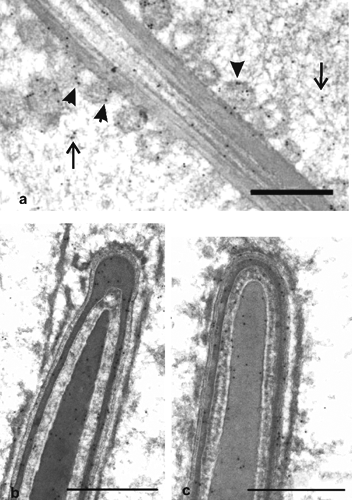
Figure 5. Immunoelectron microscopic localisation of phospholipid hydroperoxide glutathione peroxidase in spermatogenic cells of bull testis: elongated spermatids. Labelling appears in the matrix and the outer membrane of mitochondria (a, arrows), in residual cytoplasm (a, arrow-heads), and the outer dense fibres of the midpiece (a). Signals are also observed in the condensed chromatin, nuclear envelope and acrosome (b,c). Bars=0.5 μ.
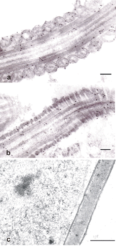
Figure 6. Immunogold labelling of phospholipid hydroperoxide glutathione peroxidase in ejaculated spermatozoa. Signals are associated with the outer mitochondrial membrane and outer dense fibres surrounding the flagellar axoneme (a). Immunocytochemical response is observed in the outer dense fibres and fibrous sheet of the main piece of the tail (b). Gold particles are visible in the cytoplasmic droplets of spermatozoa (c). Bars a,b=0.1 μ; c=0.5 μ.

Table 1. Localisation of phospholipid hydroperoxide glutathione peroxidase immunosignals at light microscopy.