Figures & data
Reproduced from Citation[106].
![Figure 1. Structural organization of cellulose from individual molecules to crystal, microfibrils, plant cell walls and finally whole plants.Reproduced from Citation[106].](/cms/asset/e3f5f855-72f6-4f72-bd87-17bf550658d2/tbfu_a_10816103_f0001.gif)
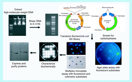
DNA is prepared, sheared, ligated into a vector, screened for positive colonies, and then the protein is expressed and characterized.
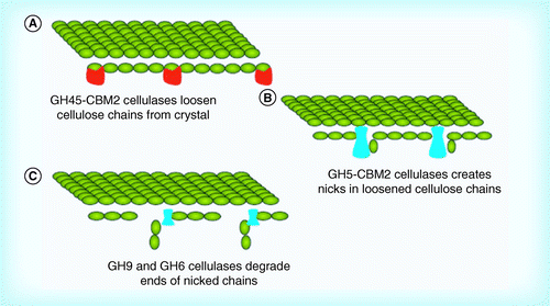
(A) A GH45 decrystallization of cellulose; (B) nicking of decrystallized cellulose; and (C) degradation of nicked chains.
CBM: Carbohydrate binding modules; GH: Glycosyl hydrolase.
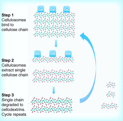
(A) Binding of cellulose to outer membrane; (B) abstraction of a single cellulose chain; (C) translocation of chain through outer membrane pore; and (D) cleavage of chain in periplasm and transport of oligosaccharides through inner membrane. Based on alginate transport and utilization model Citation[85].
![Figure 5. Potential mechanism for decrystallization and cellulose hydrolysis by Fibrobacter succinogenes, Cytophaga hutchinsonii and Sorangium cellulosum. (A) Binding of cellulose to outer membrane; (B) abstraction of a single cellulose chain; (C) translocation of chain through outer membrane pore; and (D) cleavage of chain in periplasm and transport of oligosaccharides through inner membrane. Based on alginate transport and utilization model Citation[85].](/cms/asset/f6a0cc3e-6a74-45ed-9dbd-a04b13de24f5/tbfu_a_10816103_f0005.gif)
Table 1. Potential cellulases of Gram-positive organisms.
Table 2. Cellulases of Clostridiaceae.
Table 3. Cellulases of Gram-negative organisms.