Figures & data
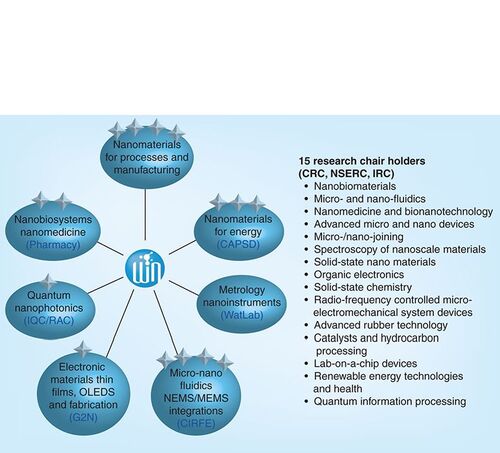
CRC: Canada Research Chair; IRC: Industrial Research Chair; NSERC: Natural Sciences and Engineering Research Council.
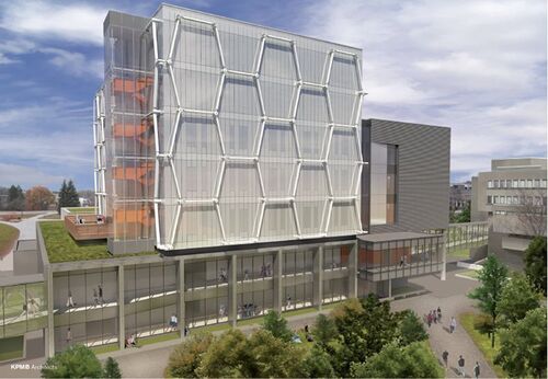
Reproduced with permission from KPMB Architects.
(A)‘Naked’ plasmid. (B) Initial phases of compaction 30 s after adding gemini surfactant; bar 100 nm. (C) Nanoparticle formation 15 min after adding gemini surfactant; bars 400 nm.
Reproduced with permission from Citation[12].
![Figure 3. Atomic-force microscopic images of the compaction of plasmid DNA into nanoparticles by gemini surfactant 16–3–16. (A)‘Naked’ plasmid. (B) Initial phases of compaction 30 s after adding gemini surfactant; bar 100 nm. (C) Nanoparticle formation 15 min after adding gemini surfactant; bars 400 nm.Reproduced with permission from Citation[12].](/cms/asset/2614c690-1e70-4804-9770-468f966c3270/ifmc_a_12362343_f0003.jpg)
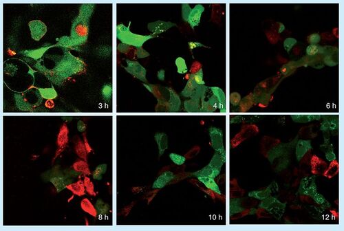
Cell viability is indicated by calcein staining (green); siGLO-RNA uptake into cells is indicated by red.
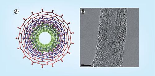
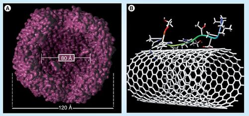
(A) Visualized internal cavity of bacterioferritin showing the nanoscale dimension of this class of protein. (B) Peptide–single-walled nanotube interaction based on various biophysical studies conducted by the Honek group.
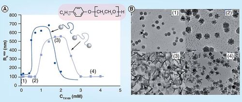
(A) CTX100 = 0 mM; (B) CTX100 = 0.7 mM; (C) CTX100 = 1.7 mM; (D) CTX100 = 3.5 mM.