Figures & data
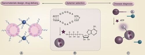
(B) Selection of aptamers for metabolite binding. An ATP aptamer and its target binding is shown. (A) Selected aptamers can be used for assembly of advanced nanomaterials such as liposome–gold nanoparticle hybrids. (C) Aptamers can also be made into highly sensitive biosensors.
F: Fluorophore; MMP: Magnetic microparticle; Q: Quencher.
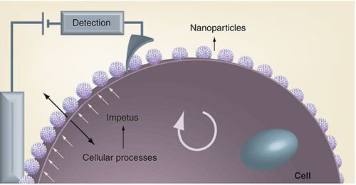
The cellular processes provide an impetus to the nanoparticles in the form of electrochemical modulation. The electronic interface, with a nanotip scanning electrode allows localized measurement of the cellular processes.
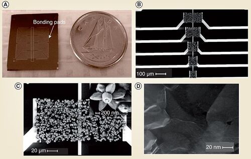
(A) Optical image of a chip compared with a Canadian dime. (B) SEM image shows a set of devices on the chip with nanostructures bridging macroscopic metal electrodes. (C) and (D) Zoomed in SEM images of flower-like ZnO nanostructures and graphene, respectively.
SEM: Scanning electron microscopy.
(A) Principle of atomic force microscopy. (B) Atomic-force microscopy image of amyloid-β1–42 fibrils
(A) With permission from Citation[101] © JPK instruments.
(B) With permission from Choi Y and Leonenko Z (Leonenko’s group) [Unpublished Data].
![Figure 4. Principle of atomic force microscopy and an example atomic-force microscopy image. (A) Principle of atomic force microscopy. (B) Atomic-force microscopy image of amyloid-β1–42 fibrils(A) With permission from Citation[101] © JPK instruments.(B) With permission from Choi Y and Leonenko Z (Leonenko’s group) [Unpublished Data].](/cms/asset/0ba9f77a-0551-428e-a7fc-c132aef69bf5/ifmc_a_12362357_f0004.jpg)
(A) Atomic-force microscopy images, (B) Kelvin force probe microscopy images and (C) Cross-section plot, animal lipid-extract surfactant film (bles) supported on mica.
Reprinted with permission from Citation[13] © Elsevier.
![Figure 5. Images obtained by atomic-force microscopy approaches (A) Atomic-force microscopy images, (B) Kelvin force probe microscopy images and (C) Cross-section plot, animal lipid-extract surfactant film (bles) supported on mica.Reprinted with permission from Citation[13] © Elsevier.](/cms/asset/e8da9ed7-b0e5-4b99-a59a-1798cdcc89e4/ifmc_a_12362357_f0005.jpg)