Figures & data
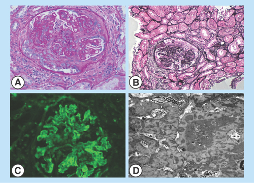
Figure 1. Renal biopsy images.
(A&B) Light microscopy of sections of renal cortex and medulla with active cellular and fibrocellular crescents. Frequent spike formation and segmental subendothelial deposits are seen. (C) Direct immunofluorescence of the renal cortex shows glomeruli with granular staining in the mesangial and capillary walls for IgG, IgA, IgM, C3, C1q, kappa and lambda light chains. (D) Electron microscopy of a glomerulus exhibiting immune-type electron-dense deposits in the subepithelial, intramembranous and mesangial locations. Glomerular basement membranes are irregularly thickened and distorted by the deposits.