Figures & data
Figure 1. MRI of knee osteoarthritis showing bone marrow lesion.
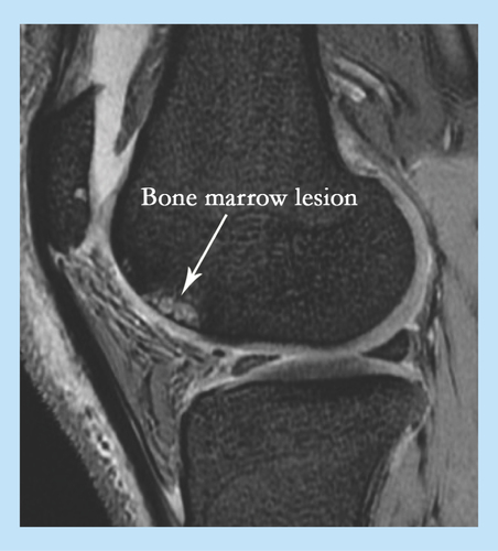
Figure 2. Bone remodeling process.
(A) Signals involved in bone remodeling and the role of mesenchymal stem cells (MSCs) in these processes. MSCs are precursors of osteoblasts (black arrow). The process of osteoblastogenesis is inhibited by sclerostin produced by osteocytes. In concert with osteocytes, MSCs also control osteoclastogenesis via the production of RANKL and OPG (blue arrow). In OA, osteoclasts release TGF-β from the bone matrix, which modulates MSC activity near the resorption site. (B) Bone remodeling activity in OA subchondral bone (hematoxylin and eosin staining of EDTA-decalcified femoral head subchondral bone). OA: Osteoarthritis.
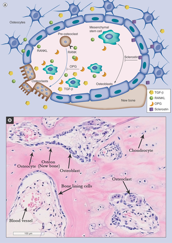
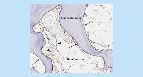
Figure 3. Mesenchymal stem cell appearance in osteoarthritis subchondral bone showing CD271+ staining in perivascular (arrow heads) and bone lining (arrows) locations particularly in the areas of newly-formed bone.