Figures & data
Figure 1. MCF-12A cell embedded in Matrigel gradually form acini-like structures.
Optical microscopy images of MCF-12A. (A) Cells appeared in small clusters after 24 h. They gradually formed acini-like spheroids after 4 days (B), which grow in size after 10 days (C). Fourteen days after being embedded in Matrigel, spheroids preserved their size and some of them were more defined, with clear edges (D; arrowheads), while others were less defined (D; insert, arrows). Scalebars: 250 μm.
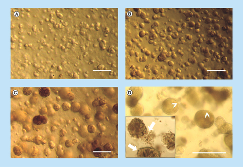
Figure 2. Hybri-Care culture medium promotes the formation of more defined acini-like structure than Dulbecco's modified Eagle medium/F12 culture medium.
Optical microscopy images of MCF-12A cell embedded in Matrigel after 14 days of culture. When using Dulbecco's modified Eagle medium/F12 medium to maintain cultures, resulting multicellular structures seem smaller, less defined and looser (A–C, arrowheads) than cells maintained in Hybri-Care medium (D–F). In Hybri-Care medium, acini are bigger, rounder, more defined and more compact. Scalebars: 250 μm.
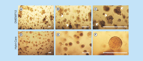
Figure 3. Presence of a lumen-like cavity in the center of cryosectioned MCF-12A acini.
Confocal microscopy images of acini immunolabeled with an E-cadherin (adherens junctions)-specific antibody (green). Nuclei are stained with 4′,6-diamidino-2-phenylindole (blue). (C) Optical microscopy image of acini cryosection stained with Masson's trichrome staining. (D) Confocal microscopy image of an acini cryosection immunolabeled with a β-catenin (adherens junctions)-specific antibody (white). Nuclei are stained with 4′,6-diamidino-2-phenylindole. Scalebars: 100 μm.
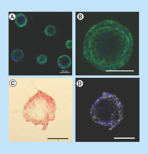
Figure 4. Matrigel at a concentration of 75% is optimal for 3D culture of MCF-12A cells.
Representative single confocal images (A, C & E) or serial Z-stacks (B, D & F) of MCF-12A cells embedded in 50% (A & B), 75% (C & D) or 100% (E & F) Matrigel and immunolabeled with an E-cadherin antibody (green). Nuclei are stained with 4′,6-diamidino-2-phenylindole (blue). Scalebars: 100 μm.
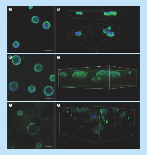
Figure 5. MCF-12A cells co-cultured with Hs578Bst cells produce acini-like structures when embedded in Matrigel.
(A & B) Representative optical microscopy images of MCF-12A cells embedded in Matrigel. (C & D) Optical microscopy images of MCF-12A cells co-cultured with Hs 578Bst cells showing acini-like structures. A second layer of cells seems to be surrounding the first layer in some of the acini (arrowhead), suggesting that myoepithelial cells formed bilayered acini with the luminal cells. (E) Optical microscopy image of Hs 578Bst cells embedded in Matrigel. Scalebars: 100 μm.
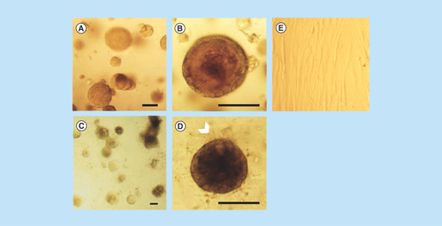
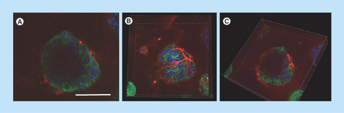
Figure 6. MCF-12A and Hs 578Bst co-cultured cells form bilayered acini in Matrigel.
(A–C) Confocal microscopy images of MCF-12A and Hs 578Bst cells forming a 3D bilayered acini when co-cultured in Matrigel. Acini were immunolabeled with an E-cadherin-specific antibody (green) and an smooth muscle actin-specific antibody (red). Nuclei are stained with 4′,6-diamidino-2-phenylindole (blue). Representative single confocal image (A), serial Z-stack (B) and a truncated view of a Z-strac (C). The myoepithelial-like cells Hs 578Bst have a stellate phenotype, forming a discontinuous basket-like network around the MCF-12A luminal cells. The truncated view suggests the presence of a lumen in the center of the bilayered acini. Scalebar: 100 μm.