Figures & data
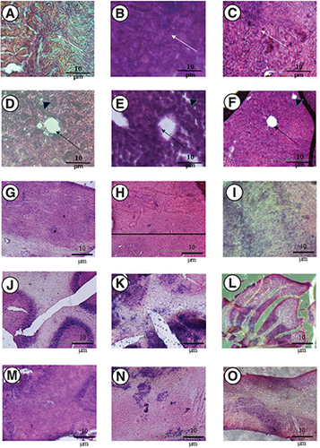
(A–O) Three groups of mice were used including, a negative control group; an intravenous group (injected with 4 mg/kg of Amphotericin B deoxycholate, referred to as iv.); and an intranasal group (2 mg/kg) + iv. group (2 mg/kg, referred to as IN group). Note that A (negative control), and C (IN group) show that glomeruli are devoid of ischemia and integrity of renal tubules remain unscathed with no signs of necrosis, while group B (iv.) exhibited tubular necrosis (white arrow). The sagittal sections of a liver lobular unit from caudate lobes are shown for (D) negative control; (E) iv. group; (F) IN group. Note that the architecture of the liver lobular unit is normal for D (negative control), and F (IN group), whereas the hepatocyte shredded into the hepatic central vein in group E (iv. group) mice (black arrow) and the intrahepatic duct was distorted with malalignment of hepatic acinus (black arrowhead). The tissues were stained in haematoxylin-eosin and were visualized under an inverted microscope (x400). (G–O) Shows different regions of the brain. For the olfactory bulb, (G) negative control; (H) iv. group; and (I) IN group. For the cerebellum, (J) negative control; (K) iv. group (L) IN group. For regions of pons – midbrain (M) negative control; (N) iv. group; (O) IN group.