Figures & data
Figure 1. CT scan of the abdomen performed before the start of epirubicin plus dacarbazine treatment and 6 months later. The hepatic and adenopathic lesions regressed from 70.3 mm (A) to 36.8 mm (B) and from 58.9 × 45 mm (C) to 21.6 × 29.5 mm respectively (D).
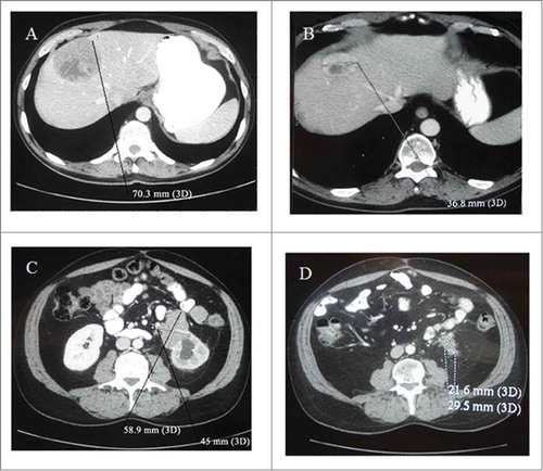
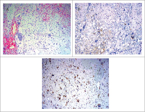
Figure 2. Microscopic features of the grade 4 unclassified renal cell carcinoma, with sarcomatoid component (URCCSC). (A) Patternless arrangement of tumor cells with pleomorphic and spindle cell morphology (H&E x10). (B) Focal immunopositivity for Keratins of neoplastic cells, showing epithelial differentiation. panCK (AE1, AE3,PCK26 clone) (H&E x 25). (C) Immunopositivity for S-100 of neoplastic cells. S-100 clone (H&E × 25).