Figures & data
Table 1. HMCLs' characteristics and sensitivity to curcumin
Figure 1. Curcumin induced cell death of myeloma cells belonging to the main molecular myeloma subgroups. (A) HMCL (n = 29) were treated with curcumin for 24 h. Cell death was measured by FACS analysis of Apo2.7 stained cells. LD50 values were calculated from at least 3 independent experiments. HMCLs were classified according to translocation subtypes. Statistical analysis was performed using Kruskall-Wallis test. Curcumin LD50 of HMCLs were analyzed according to (B) the presence or absence of t(11,14) translocation () or (C) TP53 status (). Statistical analysis was performed using Mann-Whitney test.
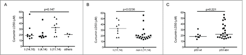
Table 2. Sensitivity of primary myeloma cells to curcumin
Figure 2. Primary myeloma cells were killed by curcumin. Primary cells (CD 138+) obtained from sample 4 were treated 24 h with the indicated doses of curcumin. Cells were then stained with an anti-CD138-PE mAb. The loss of CD138 staining was representative of cell death. Cell death percentage was calculated relative to untreated cells.
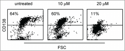
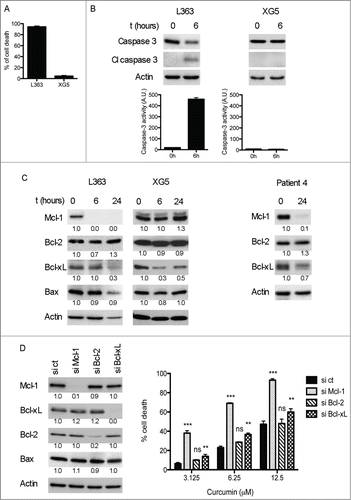
Figure 3. Curcumin cell death was associated with Mcl-1 decrease and Caspase-3 activation. (A) L363 (highly sensitive) and XG5 (poorly sensitive) cells were treated during 24 h with 15 μM curcumin, cell death was determined by Apo 2.7 staining. Data represent the mean ± SD of 3 independent experiments. (B) Caspase-3 protein levels and activity were determined on cell lysates. Data represent mean ± SD of 3 independent experiments. (C) Cells were treated with curcumin for the indicated times and the expression of Bcl-2 family molecules was assessed by protein gel blotting. Primary purified CD138+ cells (p4 sample) were treated 24 h with 15 μM curcumin, which induced 82% of cell death. (D) Transient knock-down of Mcl-1, Bcl-2 and Bcl-xL was performed on LP1 cells. After 48 h of transfection cells were treated with curcumin for the next 24 h and subjected to Apo 2.7 staining. Efficient silencing of the 3 anti-apoptotic proteins is shown by immunoblotting in the left panel. Data represent mean ± SD of 3 independent experiments. *** = P < 0.001; ** = P < 0.01; * = P < 0.05 and ns = P > 0.05. Statistical analysis was performed using 2-way Anova test.