Figures & data
Figure 1. Functional compartmentalization of liver: periportal hepatocytes on left and perivenous hepatocytes on right, with arrow in direction of blood flow.
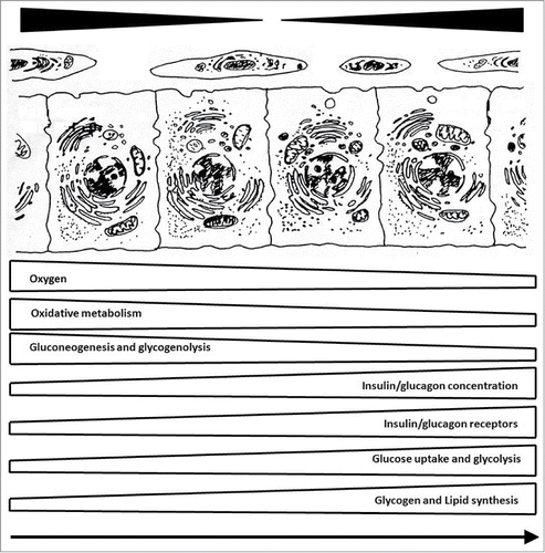
Table 1A. Zonation of cells, receptors, metabolism and biotransformation in liver. Key: +++ predominant localization in periportal or perivenous zone.
Table 1B. Zonation of enzyme activity and protein synthesis in liver. Key: +++ predominant localization in periportal or perivenous zone.
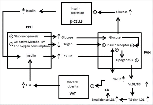
Figure 2. Relationship between visceral obesity, insulin resistance, functional compartmentalization and liver steatosis. Visceral adipocytes (VAT), periportal hepatocytes (PPH), perivenous hepatocytes (PVH) and β cells of pancreas (β-CELLS) are represented by squares. Key:↑augmentation; ⊕ stimulation; cardiovascular disease (CV); low-density lipoproteins (LDL); very low density lipoprotein (VLDL); triglyceride (TG).