Figures & data
Table 1. Protein expression of αA- and αB-crystallin in the retina of 20-day-old and 4- and 24-month-old senescence-accelerated OXYS rats
Figure 1. Treatment with 250 nmol/kg per day of SkQ1, starting at 1.5 months of age, attenuated the development of retinopathy in OXYS rats. The data are presented as stages (0, 1, 2, and 3) of retinopathy in 4-month-old control (untreated) and SkQ1-treated OXYS rats. In each group, 50 eyes of 25 animals were examined.
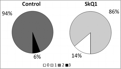
Table 2. Morphometric parameters of the chorioretinal complex of 4-month-old Wistar and OXYS rats and SkQ1-treated OXYS rats
Figure 2. The morphology of the retina of 4-month-old rats. (A) In Wistar rats, RPE cells had a prismatic shape with oval nuclei (black arrows), normal retinal layers. (B) In OXYS rats, stasis, and sludge of the blood cells are visible in capillaries of the choroid, RPE cells were flat, with a variable size and shape of the nuclei (black arrows), pyknosis of nuclei of neurosensory cells. (C and D) In OXYS rats: treatment with SkQ1 prevented anomalies in the RPE cells (black arrows) and decreased the number of neurosensory cells with pyknotic nuclei in the outer and in the inner nuclear layer. The scale bar: 10 μm, staining: H&E; abbreviations: outer nuclear layer (ONL), outer plexiform layer (OPL), inner nuclear layer (INL), inner plexiform layer (IPL), and retinal pigment epithelium (RPE).
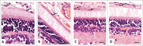
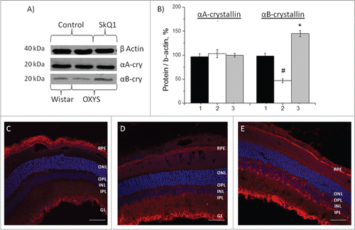
Figure 3. Effects of treatment with SkQ1 on the protein levels of αА- and αВ-crystallin in the retina of OXYS rats. (A) Western blot analysis. (B) The protein gel blot results quantified as a percent of data from untreated age-matched Wistar rats (mean ± SEM), normalized to β-actin from 5 independent experiments. 1: Wistar rats, 2: OXYS rats, 3: OXYS rats treated with SkQ1. #Significant differences between untreated OXYS and Wistar rats (P < 0.05); *a significant effect of treatment with SkQ1 (P < 0.05). Confocal immunofluorescent images depict αB-crystallin (red signal) detected within the retina and RPE in OXYS rats (C) compared to disease-free Wistar (D) rats and (E) SkQ1-treated OXYS rats (250 nmol/kg per day from 1.5 to 4 months of age). Cell nuclei were stained with DAPI (blue). The scale bar: 50 μm. Abbreviations: outer nuclear layer (ONL), outer plexiform layer (OPL), inner nuclear layer (INL), inner plexiform layer (IPL), retinal pigment epithelium (RPE), and ganglion layer (GL).