Figures & data
Figure 1. MFCM dedifferentiate differentiated cells to CSCs Spheroid cultures grown in control medium (red) or MFCM (brown) for 24 h and were (A) stained with stem cell marker Lgr5, CD133 or (B) qrt-pcr was performed on differentiation markers Ck20 and Mucin2 (Muc2). (C) Limiting dilutions experiments were performed and clonogenic fraction was calculated in CSC (TOP-GFPhi), Differentiated cell (TOP-GFPlo), and differentiated cells deposited in MFCM (Diff + MFCM). Significance is shown as ***P < 0.001.
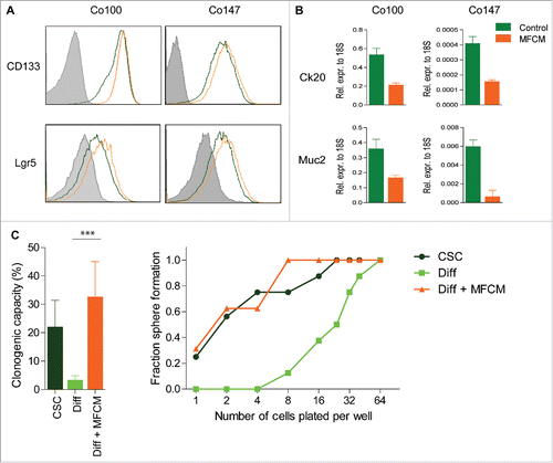
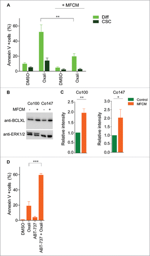
Figure 2. BCLXL is required for MFCM induced resistance toward oxaliplatin (A) Primary spheroid culture transduced with TOP-GFP construct (Co100) was exposed to MFCM or control medium for 24 h. Subsequently, cells were treated for 24 h with oxaliplatin and Annexin V/ 7AAD staining was performed. CSCs and differentiated cells were defined by gating on TOP-GFPhi and TOP-GFPlo expressing cells, respectively. MFCM treatment decreases oxaliplatin induced cell death. (B) Western blot analysis of BCLXL protein in spheroid cultures grown in control or MFCM for 24 h. Increased BCLXL expression in spheroid culture treated with MFCM compared to control medium. Control western blots for ERK1/2 is shown in lower panel. (C) Quantification of BCLXL protein intensity relative to ERK1/2 protein expression. Average of 3 independent experiment is shown. (D) Co100 spheroid culture was exposed to MFCM for 24 h. Cells were treated with 100 nM ABT-737 in combination with or without oxaliplatin. Cell death was measured with AnnexinV/7-AAD staining in TOP-GFPlo cells. Significance is indicated (*P < 0.05, **P < 0.01, ***P < 0.001).