1,218
Views
7
CrossRef citations to date
0
Altmetric
REPORTS

The alternative translated MDMXp60 isoform regulates MDM2 activity

, , , , &
Pages 449-458 | Received 04 Jul 2014, Accepted 12 Oct 2014, Published online: 06 Feb 2015

Figures & data

Figure 1. The hMDMXp60 isoform is initiated at the 7th in frame AUG codon. (A) Cartoon illustrating the hmdmx mRNA constructs and the mutated AUG sites (left). The 6 in frame AUG codons after the +1 AUG of the hmdmx coding sequence were substituted with alanine (GCG) (hmdmxΔ2–7AUG) to express only full length hMDMX (hMDMXFL). The hmdmxΔ1–6AUG mRNA lacks the first 6 in frame AUG codons and only expresses hMDMXp60 which is initiated from the 7th in frame AUG (+384). The hMDMXp60 lacks the first 127 amino acids, including the p53 binding domain (right). (B) Western blot showing the expression of the two hMDMX isoforms from indicated constructs in H1299 cells. The hmdmxwt mRNA expresses both isoforms. Actin is used as loading control. (C) The relative expression of the two hMDMX isoforms in different cell lines. H1299 and Saos-2 are p53 negative cell lines whereas HeLa expresses wild type p53 and MDA-MB-231 expresses a mutant p53(R280K). Western blots show one representative experiment out of 3.
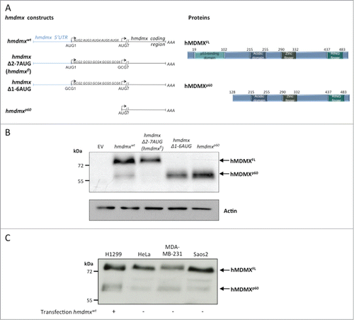
Figure 1. The hMDMXp60 isoform is initiated at the 7th in frame AUG codon. (A) Cartoon illustrating the hmdmx mRNA constructs and the mutated AUG sites (left). The 6 in frame AUG codons after the +1 AUG of the hmdmx coding sequence were substituted with alanine (GCG) (hmdmxΔ2–7AUG) to express only full length hMDMX (hMDMXFL). The hmdmxΔ1–6AUG mRNA lacks the first 6 in frame AUG codons and only expresses hMDMXp60 which is initiated from the 7th in frame AUG (+384). The hMDMXp60 lacks the first 127 amino acids, including the p53 binding domain (right). (B) Western blot showing the expression of the two hMDMX isoforms from indicated constructs in H1299 cells. The hmdmxwt mRNA expresses both isoforms. Actin is used as loading control. (C) The relative expression of the two hMDMX isoforms in different cell lines. H1299 and Saos-2 are p53 negative cell lines whereas HeLa expresses wild type p53 and MDA-MB-231 expresses a mutant p53(R280K). Western blots show one representative experiment out of 3.

Figure 2. (See previous page) hMDMXp60 and hMDMXFL are derived from cap-independent mRNA translation initiation. (A) Cartoon illustrating the bicistronic hmdmx mRNA inserted downstream of the GFP open reading frame and downstream of a hairpin structure preventing ribosomal read through from cap-dependent translation initiation of the GFP open reading frame. The location of primers (P1 to P5) is indicated. These were used for RT-PCR and RT-qPCR to ensure that the bicistronic mRNA remains intact in cells and that no cryptic promoter activity causes alternative RNA species. (B) The expression of the two hMDMX isoforms from the wild type mRNA and from the bicistronic construct in H1299 cells. The relative level of expression of the two hMDMX isoforms from wild type vs. bicistronic constructs is estimated (upper graph). (C) RT-PCR using indicated primer pairs repeated 4 times each for the bicistroninc hmdmx mRNA. (D) The relative amount of RT-PCR products from the bicistronic and the wild type hmdmx mRNAs as estimated using quantitative RT-PCR from indicated primers. The ratio of RT-PCR products derived from the primers covering the 5’UTR and the initiation site for hmdmxFL (P2 and P3) and from a sequence covering the initiation site of hMDMXp60 (P4 and P5) are similar between the wild type and the bicistronic hmdmx mRNAs. This shows that no cryptic promoter or alternative splicing was created by fusing the hmdmx mRNA to the bicistronic construct. No added RT enzyme (-RT) shows that the quantified PCR products are derived from mRNA. Actin mRNA serves as control. The graph shows data from 3 independent experiments plus SD. (E) The use of siRNA against the GFP results in a similar reduction in expression of GFP and hMDMX from the bicistronic hmdmx mRNA. There is no effect on hMDMX expression form the wild type mRNA (Fig. S1). (F) Western blot showing the effect of indicated UV doses 4 hours after treatment on cap-independent translation of the hmdmx mRNA as compared to cap-dependent translation of gfp (Fig. S2). Western blots show one representative experiment out of 3.
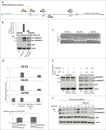
Figure 2. (See previous page) hMDMXp60 and hMDMXFL are derived from cap-independent mRNA translation initiation. (A) Cartoon illustrating the bicistronic hmdmx mRNA inserted downstream of the GFP open reading frame and downstream of a hairpin structure preventing ribosomal read through from cap-dependent translation initiation of the GFP open reading frame. The location of primers (P1 to P5) is indicated. These were used for RT-PCR and RT-qPCR to ensure that the bicistronic mRNA remains intact in cells and that no cryptic promoter activity causes alternative RNA species. (B) The expression of the two hMDMX isoforms from the wild type mRNA and from the bicistronic construct in H1299 cells. The relative level of expression of the two hMDMX isoforms from wild type vs. bicistronic constructs is estimated (upper graph). (C) RT-PCR using indicated primer pairs repeated 4 times each for the bicistroninc hmdmx mRNA. (D) The relative amount of RT-PCR products from the bicistronic and the wild type hmdmx mRNAs as estimated using quantitative RT-PCR from indicated primers. The ratio of RT-PCR products derived from the primers covering the 5’UTR and the initiation site for hmdmxFL (P2 and P3) and from a sequence covering the initiation site of hMDMXp60 (P4 and P5) are similar between the wild type and the bicistronic hmdmx mRNAs. This shows that no cryptic promoter or alternative splicing was created by fusing the hmdmx mRNA to the bicistronic construct. No added RT enzyme (-RT) shows that the quantified PCR products are derived from mRNA. Actin mRNA serves as control. The graph shows data from 3 independent experiments plus SD. (E) The use of siRNA against the GFP results in a similar reduction in expression of GFP and hMDMX from the bicistronic hmdmx mRNA. There is no effect on hMDMX expression form the wild type mRNA (Fig. S1). (F) Western blot showing the effect of indicated UV doses 4 hours after treatment on cap-independent translation of the hmdmx mRNA as compared to cap-dependent translation of gfp (Fig. S2). Western blots show one representative experiment out of 3.

Figure 3. hMDMXp60 shows higher affinity toward hMDM2 as compared to hMDMXFL. (A) Immunoprecipitation (IP) against hMDM2 shows that hMDM2 interacts with both hMDMX isoforms (upper panel). The protein levels in whole lysates are shown below. (NS stands for non-specific). (B) Co-immunoprecipitation of hMDMX isoforms (left) or hMDM2 (right) using an HA-tagged hMDMXp60. Hsp90 and actin serves as loading controls of total lysates. (C) ELISA using a fixed (500 ng) amount of recombinant purified hMDMXp60 or hMDMXFL and increasing amounts of hMDM2. The affinity of hMDMXp60 to hMDM2 is higher as compared to hMDMXFL. Both proteins bind hMDM2 in a biphasic fashion. The data shows the average of 7 independent experiments and SD. Western blots (A and B) show one representative experiment out of 3.
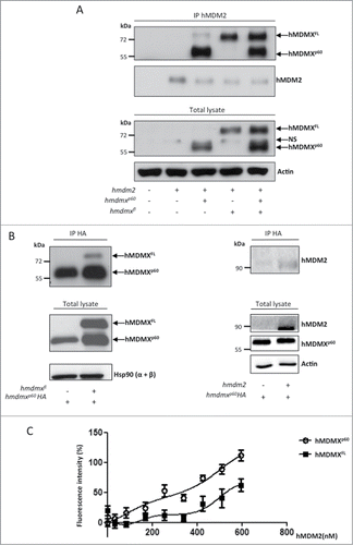
Figure 3. hMDMXp60 shows higher affinity toward hMDM2 as compared to hMDMXFL. (A) Immunoprecipitation (IP) against hMDM2 shows that hMDM2 interacts with both hMDMX isoforms (upper panel). The protein levels in whole lysates are shown below. (NS stands for non-specific). (B) Co-immunoprecipitation of hMDMX isoforms (left) or hMDM2 (right) using an HA-tagged hMDMXp60. Hsp90 and actin serves as loading controls of total lysates. (C) ELISA using a fixed (500 ng) amount of recombinant purified hMDMXp60 or hMDMXFL and increasing amounts of hMDM2. The affinity of hMDMXp60 to hMDM2 is higher as compared to hMDMXFL. Both proteins bind hMDM2 in a biphasic fashion. The data shows the average of 7 independent experiments and SD. Western blots (A and B) show one representative experiment out of 3.

Figure 4. hMDMXp60 stabilizes hMDMXFL in the presence of hMDM2. (A) The levels of expression of hMDMXFL in H1299 cells following increasing amounts of hMDM2 (left). A similar experiment but using a fixed amount of hMDMXp60 and increasing levels of hMDM2 (right). Compare differences in the expression of hMDMX isoform in cells transfected with 50 ng hmdm2 cDNA. (B) hMDMXFL is protected from hMDM2-mediated degradation in the presence of hMDMXp60. (C) Increasing levels of hMDMXp60 stabilizes hMDMXFL in cells expressing endogenous hMDM2. (D) Mutation of each, or all 6 together, of the lysine residues in the N-terminus of hMDMX upstream of the initiation site for hMDMXp60 do not affect hMDM2-mediated degradation of hMDMX. The data are representative from 3 independent experiments (Fig. S3A and B).
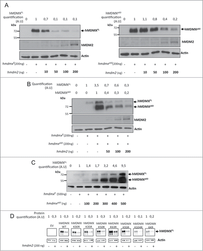
Figure 4. hMDMXp60 stabilizes hMDMXFL in the presence of hMDM2. (A) The levels of expression of hMDMXFL in H1299 cells following increasing amounts of hMDM2 (left). A similar experiment but using a fixed amount of hMDMXp60 and increasing levels of hMDM2 (right). Compare differences in the expression of hMDMX isoform in cells transfected with 50 ng hmdm2 cDNA. (B) hMDMXFL is protected from hMDM2-mediated degradation in the presence of hMDMXp60. (C) Increasing levels of hMDMXp60 stabilizes hMDMXFL in cells expressing endogenous hMDM2. (D) Mutation of each, or all 6 together, of the lysine residues in the N-terminus of hMDMX upstream of the initiation site for hMDMXp60 do not affect hMDM2-mediated degradation of hMDMX. The data are representative from 3 independent experiments (Fig. S3A and B).

Figure 5. Increasing levels of hMDMXp60 induces oscillation in hMDM2 expression levels in H1299 cells. (A) Expression of a fixed amount of hmdm2 and increasing levels of either (a) hMDMXp60 alone or (b) together with a fixed amount of hmdmxfl, or (c) increasing levels of both isoforms. The hmdm2(C464A) mutant (d) is E3 ligase dead and is not affected by hMDMXp60 (Fig. S4A and B). (B) In vitro autoubiquitination of recombinant hMDM2 in the presence of increasing levels of hMDMXp60. Data shows one representative experiment out of 3 (Fig. S5A and B).
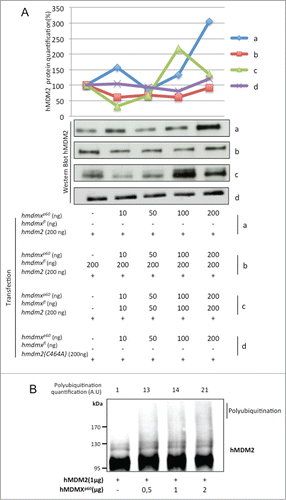
Figure 5. Increasing levels of hMDMXp60 induces oscillation in hMDM2 expression levels in H1299 cells. (A) Expression of a fixed amount of hmdm2 and increasing levels of either (a) hMDMXp60 alone or (b) together with a fixed amount of hmdmxfl, or (c) increasing levels of both isoforms. The hmdm2(C464A) mutant (d) is E3 ligase dead and is not affected by hMDMXp60 (Fig. S4A and B). (B) In vitro autoubiquitination of recombinant hMDM2 in the presence of increasing levels of hMDMXp60. Data shows one representative experiment out of 3 (Fig. S5A and B).
Supplemental material

977081_Supplementary_Materials.zip

Download Zip (21.3 MB)