
Figures & data
Figure 1. Dovitinib inhibited the breast cancer invasion and antagonize the invasion promoting-effect of CAFs. (A) One example of isolated CAFs from patient samples (B) Enhanced invasion ability of breast cancer cells MCF-7, BT-474 and MDA-MB-231 through co-culture with CAFs. Human breast cancer CAFs were seeded in 24-well-plate and cultured in serum-free medium for 3 d Breast cancer cells suspended in serum-free media were added into the inserts either with CAFs or with only serum-free medium in the bottom chamber. Invasion assay was performed as described in the Materials and Methods. Non-invaded cells were removed from the top surface of the insert by scrubbing with cotton tip swabs. 18 h later, the membranes of the inserts with invaded cells were fixed, stained, mounted on slides, and counted under light microscope. (C) Dose-dependently inhibited invasion ability of MDA-MB-231 cells after treatment with Dovitinib. Breast cancer cells MDA-MB-231 were pre-treated with Dovitinib (0.01, 0.1, 0.5 μM) for 2 days, suspended in cell culture medium, and added into the inserts with cell culture medium in the bottom chamber. Invasion assay was performed as described in the Materials and Methods. (D) Pre-treatment of MDA-MB-231 cells with Dovitinib led to inhibited invasion in the co-culture system. CAFs were seeded in 24-well-plate and cultured in serum-fee medium for 3 d Breast cancer cells MDA-MB-231 were pre-treated with Dovitinib (0.01, 0.1 μM) for 2 days, suspended in serum-free medium, and added into the inserts either with CAFs or with only serum-free medium in the bottom chamber. Invasion assay was performed as described in the Materials and Methods. (E) Pre-treatment of CAFs with Dovitinib led to inhibited invasion in the co-culture system. CAFs were seeded in 24-well-plate and pre-treated with Dovitinib (0.01 μM) for 1 day. MDA-MB-231 cells were suspended in serum-free medium, and added into the inserts either with CAFs or with only serum-free medium in the bottom chamber. Invasion assay was performed as described in the Materials and Methods.
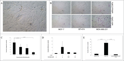
Figure 2. Application of Dovitinib in the MDA-MB-231 cells, CAFs, or the co-culture of the both types of cells resulted in the inhibited production of chemokines CCL2, CCL5 and VEGF. (A–C) 2 × 10⁴ CAFs and 2 × 10⁵ MDA-MB-231 cells were cultured alone in 24-well-plate or co-cultured with BD BioCoatTM Martrigel Invasion Chambers in serum-fee medium for 2 d in the presence or absence of Dovitinib (0.1 μM). After incubation, cell supernatants of MDA-MB-231 cells, CAFs, and co-culture were collected and served for ELISA measurement, cells were lysed for total RNA isolation. A: ELISA measurement of CCL2 and CCL5 production; B: Real-time PCR determination of CCL2 and CCL5 expression in MDA-MB-231 cells, CAFs, CAFs in co-culture with breast cancer cells; C: ELISA measurement of VEGF production.
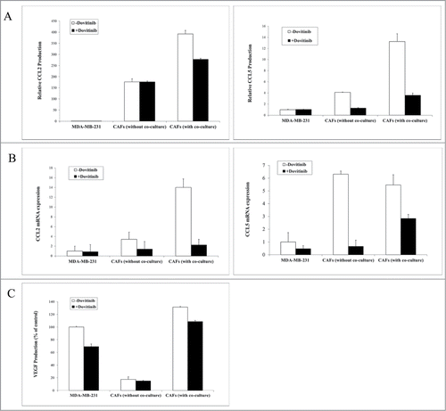
Figure 3. Dovitinib antagonized the PDGF/FGF stimulated MDA-MB-231 cell invasion and chemokine production of CAFs and MDA-MB-231 cells. (A) Dovitinib inhibited the PDGFß expression of MDA-MB-231 cells stimulated with CAFs-conditioned media.CAFs were cultured in serum-free medium for 3 d Cell culture media was collected for further experiment. MDA-MB-231 cells were cultured with serum-free medium or CAFs-conditioned media in the presence of Dovitinib (0.1 μM) for 2 d MDA-MB-231 cells were lysed and PDGFß expression was measured with real-time PCR. Each value presented is the mean ± SD of 3 parallel experiments. (B) Dovitinib antagonized the PDGF/FGF stimulated cell invasion of MDA-MB-231 cells.MDA-MB-231 cells were treated with Dovitinib (0.1 μM) for 2 d Cell invasion assay was performed in serum-free medium, serum-free medium including 10 ng/ml PDGF, or serum-free medium including 10 ng/ml FGF respectively. Each value presented is the mean ± SD of 3 parallel experiments. (C–H) Dovitinib treatment resulted in reduced CCL5, CCL2 and VEGF production in CAFs and MDA-MB-231 cells.CAFs (C-E) or MDA-MB-231 cells (F-H) were treated with PDGF (10 ng/ml) or FGF (10 ng/ml) in the presence or absence of Dovitinib (0.1 μM) for 2 d Cell supernatants were collected; CCL5, CCL2 or VEGF concentration was measured with ELISA.
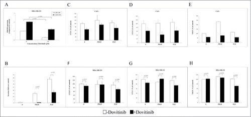
Figure 4. Dovitinib impaired Akt signaling in CAFs and led to enhanced inhibitory effect on cell invasion by combination with PI3K/Akt/mTOR signaling inhibitors Ly294002 or RAD001. (A) CAFs were cultured in serum-free RPMI-1640 media, serum-free RPMI-1640 media supplemented with 10 ng/ml PDGF, or MDA-MB-231 conditioned media (CM) in the presence or absence of Dovitinib for 24 h respectively. Total protein was extracted; the expression of phosphorylated Akt (T308) and phosphorylated Akt (S473) was determined by Western blot analysis. The relative intensity of each band shown under the band was quantified using PhoretixTM ID Quantifier software, normalized to ß-actin and expressed as percentage of control. (B) CAFs were seeded in 24-well-plate and cultured in serum-fee medium in the presence of Ly294002 (20 μM), RAD001 (10 nM), Ly294002 (20 μM)+Dovitinib (0.1 μM), or RAD001(10 nM)+Dovitinib (0.1 μM), respectively for 3 d MDA-MB-231 cells were suspended in serum-free media and added into the inserts either with or without CAFs in the bottom wells. Transwell® invasion assay was performed as described in the Materials and Methods. Each value presented is the mean ± SD of 3 parallel experiments.
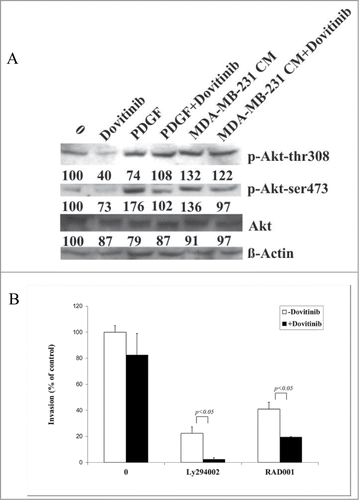
Table 1. Primers and their sequences used in the real-time PCR