998
Views
7
CrossRef citations to date
0
Altmetric
Reports

TGN38 is required for the metaphase I/anaphase I transition and asymmetric cell division during mouse oocyte meiotic maturation

, , , , , & show all
Pages 2723-2732 | Received 08 May 2014, Accepted 09 Jun 2014, Published online: 30 Oct 2014

Figures & data

Figure 1. Expression and localization of TGN38 in mouse oocytes during meiotic maturation. (A) Relative level of TGN38 mRNA in mouse oocytes during meiotic maturation. TGN38 mRNA levels were normalized to the maximum levels at GV stage. Samples were collected for quantitative RT-PCR when the oocytes were cultured for 0 h, 8 h or 12 h, corresponding to GV, MI or MII stages, respectively. Each sample contains 50 oocytes. (B) Protein level of TGN38 in mouse oocytes at GV, MI and MII stages. Each sample contains 200 oocytes. (C) Oocytes at GV, MI or MII stages were co-stained anti-TGN38 (green) and anti-γ-tubulin (red) antibodies. Besides the localization in the cytoplasm, TGN38 co-localized with γ-tubulin during oocyte maturation. Bars, 10 μm.
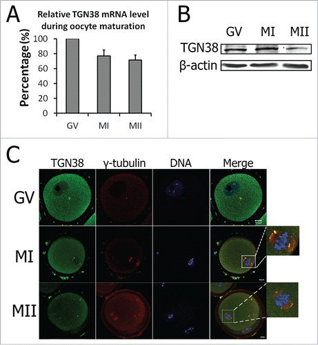
Figure 1. Expression and localization of TGN38 in mouse oocytes during meiotic maturation. (A) Relative level of TGN38 mRNA in mouse oocytes during meiotic maturation. TGN38 mRNA levels were normalized to the maximum levels at GV stage. Samples were collected for quantitative RT-PCR when the oocytes were cultured for 0 h, 8 h or 12 h, corresponding to GV, MI or MII stages, respectively. Each sample contains 50 oocytes. (B) Protein level of TGN38 in mouse oocytes at GV, MI and MII stages. Each sample contains 200 oocytes. (C) Oocytes at GV, MI or MII stages were co-stained anti-TGN38 (green) and anti-γ-tubulin (red) antibodies. Besides the localization in the cytoplasm, TGN38 co-localized with γ-tubulin during oocyte maturation. Bars, 10 μm.

Figure 2. Localization of TGN38 in mouse oocytes after treatment with nocodazole or taxol. Oocytes were incubated in M2 medium containing nocodazole (20 μg/ml; 10 min) or taxol (10 μM; 45 min). Then the oocytes were washed thoroughly with M2 medium and co-stained with antibodies against TGN38 (purple) and γ-tubulin (red). Hoechst 33342 was used to stain DNA (Blue). Bars, 10 μm.
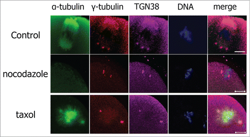
Figure 2. Localization of TGN38 in mouse oocytes after treatment with nocodazole or taxol. Oocytes were incubated in M2 medium containing nocodazole (20 μg/ml; 10 min) or taxol (10 μM; 45 min). Then the oocytes were washed thoroughly with M2 medium and co-stained with antibodies against TGN38 (purple) and γ-tubulin (red). Hoechst 33342 was used to stain DNA (Blue). Bars, 10 μm.

Figure 3. Depletion of TGN38 in mouse oocytes caused MI arrest and decreased percentage of first polar body extrusion. (A) Relative level of TGN38 mRNA in mouse oocytes after injection of scrambled siRNAs or TGN38 siRNAs. Following injection, oocytes were cultured in M2 medium containing 2.5 μM milrinone for 24 h before collected for quantitative RT-PCR. Fifty oocytes were collected in each sample. *, P < 0.001. (B) TGN38 protein level in control and TGN38 RNAi groups. Each sample contains 200 oocytes. (C) Percentages of oocytes at different stages when cultured for 12 h after TGN38 RNAi. *, P < 0.05. (D) Representative images of BubR1 localization in TGN38 depleted oocytes. Arrow: PB1. Bar, 10 μm.
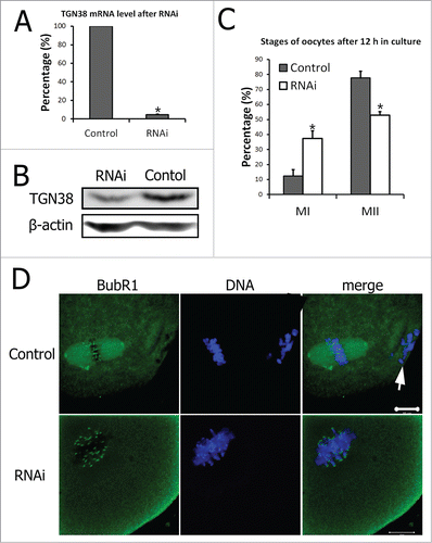
Figure 3. Depletion of TGN38 in mouse oocytes caused MI arrest and decreased percentage of first polar body extrusion. (A) Relative level of TGN38 mRNA in mouse oocytes after injection of scrambled siRNAs or TGN38 siRNAs. Following injection, oocytes were cultured in M2 medium containing 2.5 μM milrinone for 24 h before collected for quantitative RT-PCR. Fifty oocytes were collected in each sample. *, P < 0.001. (B) TGN38 protein level in control and TGN38 RNAi groups. Each sample contains 200 oocytes. (C) Percentages of oocytes at different stages when cultured for 12 h after TGN38 RNAi. *, P < 0.05. (D) Representative images of BubR1 localization in TGN38 depleted oocytes. Arrow: PB1. Bar, 10 μm.

Figure 4. TGN38 depletion caused failure of asymmetric cell division in mouse oocytes. (A) Percentages of symmetric division in oocytes when cultured for 12 h after TGN38 RNAi. Data are represented as mean ± s.e.m. of 3 independent experiments. *, P < 0.05. (B) Representative images of oocytes at 12 h of incubation. Upper lane: images from an optical microscope. Arrows: oocytes that divided symmetrically. Arrow heads: oocytes without a polar body. Lower lane: Oocytes were co-stained with anti-α-tubulin (Green) antibody and Hoechst 33342 (Blue), and examined under a confocal microscope. Bars, 10 μm.
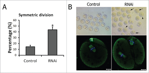
Figure 4. TGN38 depletion caused failure of asymmetric cell division in mouse oocytes. (A) Percentages of symmetric division in oocytes when cultured for 12 h after TGN38 RNAi. Data are represented as mean ± s.e.m. of 3 independent experiments. *, P < 0.05. (B) Representative images of oocytes at 12 h of incubation. Upper lane: images from an optical microscope. Arrows: oocytes that divided symmetrically. Arrow heads: oocytes without a polar body. Lower lane: Oocytes were co-stained with anti-α-tubulin (Green) antibody and Hoechst 33342 (Blue), and examined under a confocal microscope. Bars, 10 μm.

Figure 5. Peripheral spindle migration at MI stage was impaired after TGN38 depletion in mouse oocytes. (A) Representative images of oocytes in different siRNAs injected groups. After cultured for 8.5 h following TGN38 RNAi, oocytes were fixed and co-stained with anti-α-tubulin (Green) antibody and Hoechst 33342 (Blue). Bars, 10 μm. (B) Percentages of position of MI spindle in oocytes. *, P < 0.05. (C) Percentages of oocytes with abnormal spindles. *, P < 0.05.
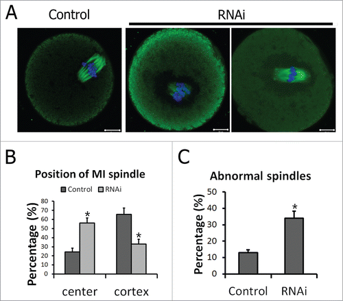
Figure 5. Peripheral spindle migration at MI stage was impaired after TGN38 depletion in mouse oocytes. (A) Representative images of oocytes in different siRNAs injected groups. After cultured for 8.5 h following TGN38 RNAi, oocytes were fixed and co-stained with anti-α-tubulin (Green) antibody and Hoechst 33342 (Blue). Bars, 10 μm. (B) Percentages of position of MI spindle in oocytes. *, P < 0.05. (C) Percentages of oocytes with abnormal spindles. *, P < 0.05.
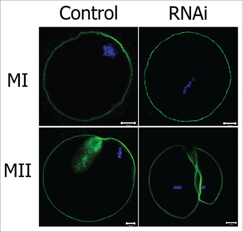
Figure 6. Effect of TGN38 depletion on actin cap formation in mouse oocytes. Oocytes were co-stained with Phalloidin-FITC (green) and Hoechst 33342 (Blue). Bars, 10 μm.

Figure 6. Effect of TGN38 depletion on actin cap formation in mouse oocytes. Oocytes were co-stained with Phalloidin-FITC (green) and Hoechst 33342 (Blue). Bars, 10 μm.

Reprints and Corporate Permissions

Please note: Selecting permissions does not provide access to the full text of the article, please see our help page How do I view content?

To request a reprint or corporate permissions for this article, please click on the relevant link below:

Academic Permissions

Please note: Selecting permissions does not provide access to the full text of the article, please see our help page How do I view content?

Obtain permissions instantly via Rightslink by clicking on the button below:

If you are unable to obtain permissions via Rightslink, please complete and submit this Permissions form. For more information, please visit our Permissions help page.