1,012
Views
1
CrossRef citations to date
0
Altmetric
BRIEF REPORT

An unusual colon atresia in a calf: at the junction of the distal loop and transverse colon. A brief overview

&
Pages 312-316 | Received 28 Jul 2014, Accepted 18 Sep 2014, Published online: 21 Jan 2015

Figures & data

Figure 1. Schematic right lateral view of the bovine large intestinal tract. 1) Cecum; Ascending colon: 2a) Proximal loop, 2b) Spiral colon, 2c) Distal loop; 3) Transverse colon; 4) Descending colon; I) Ileum; A) Aorta; CA) Celiac artery; CMA) Cranial mesenteric artery; CdMA) Caudal mesenteric artery. The bold line shows where the atresia coli was placed. Modified from ref. 29.
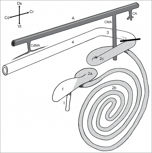
Figure 1. Schematic right lateral view of the bovine large intestinal tract. 1) Cecum; Ascending colon: 2a) Proximal loop, 2b) Spiral colon, 2c) Distal loop; 3) Transverse colon; 4) Descending colon; I) Ileum; A) Aorta; CA) Celiac artery; CMA) Cranial mesenteric artery; CdMA) Caudal mesenteric artery. The bold line shows where the atresia coli was placed. Modified from ref. 29.

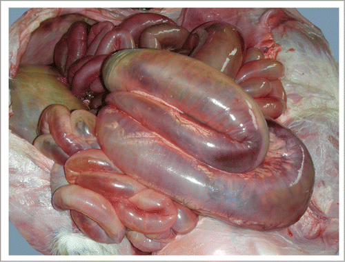
Figure 2. Dilated intestine: 1) Jejunum; 2) Cecum; 3) Spiral loop of ascending colon.

Figure 2. Dilated intestine: 1) Jejunum; 2) Cecum; 3) Spiral loop of ascending colon.

Figure 3. Distended spiral loop of the ascending colon.

Figure 3. Distended spiral loop of the ascending colon.

Figure 4. Distal loop of ascending colon showing congestion. This picture shows both blind ends of the colon: (*) proximal and (˚) distal ends.
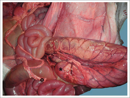
Figure 4. Distal loop of ascending colon showing congestion. This picture shows both blind ends of the colon: (*) proximal and (˚) distal ends.
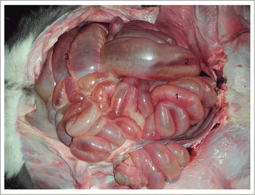
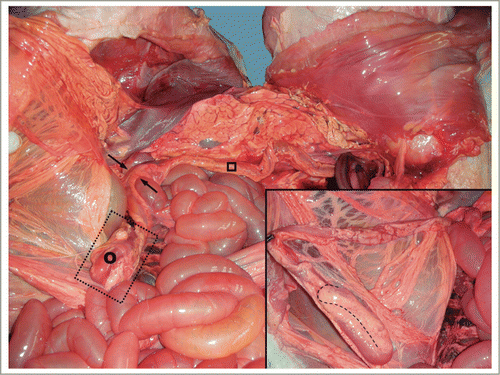
Figure 5. The distal blind end (transverse colon) was bent into a U-shape. (˚) Transverse colon; (□) descending colon; () Left colic flexure. Inset: major detail of the distal blind end.

Figure 5. The distal blind end (transverse colon) was bent into a U-shape. (˚) Transverse colon; (□) descending colon; (→) Left colic flexure. Inset: major detail of the distal blind end.

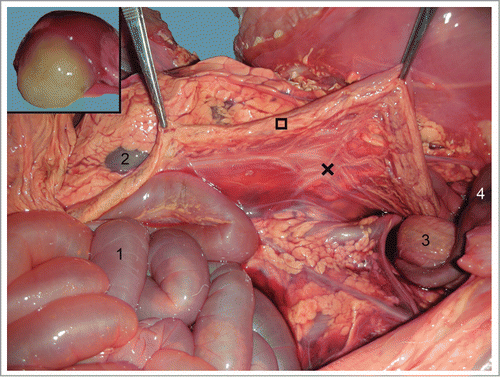
Figure 6. Descending colon (□) suspended by the mesocolon (×). 1) Jejunum; 2) Left kidney; 3) urine bladder; 4) Umbilical arteries. Inset: yellowish mucous secretion filling the descending colon lumen.

Figure 6. Descending colon (□) suspended by the mesocolon (×). 1) Jejunum; 2) Left kidney; 3) urine bladder; 4) Umbilical arteries. Inset: yellowish mucous secretion filling the descending colon lumen.

Reprints and Corporate Permissions

Please note: Selecting permissions does not provide access to the full text of the article, please see our help page How do I view content?

To request a reprint or corporate permissions for this article, please click on the relevant link below:

Academic Permissions

Please note: Selecting permissions does not provide access to the full text of the article, please see our help page How do I view content?

Obtain permissions instantly via Rightslink by clicking on the button below:

If you are unable to obtain permissions via Rightslink, please complete and submit this Permissions form. For more information, please visit our Permissions help page.