
Figures & data
Figure 1. Origin of brown, white and beige adipocytes. Two types of common precursors of muscle/fat lineage are derived from mesenchymal stem cells. These 2 types of common precursors are distinguished by Myf5, Pax7, Pax3 and PDGFRα expression. Myf5+, Pax7+ and Pax3+ common precursors give rise to specified muscle or brown adipose precursors that differentiate into either mature myocytes (Myf5+, MyoD+, Myogenin+) or brown adipocytes (UCP1+), respectively. Myf5-, Pax3-, Pax7- and PDGFRα+ common precursors give rise to specified beige or white adipose precursors that differentiate into either mature beige adipocytes (Tnfrnfs9+, Tmem26+, Tbx1+) or white adipocytes (Eva1+, Hspb7+, Pdk4+), respectively. The interconversion of mature beige and white adipocytes may occur under environmental stimulus.
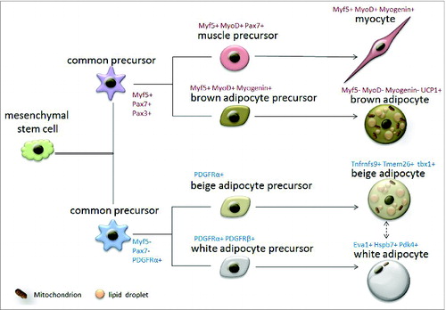
Figure 2. Distribution of various adipose depots Adipose tissues are generally divided into brown adipose depots and white adipose depots. Brown adipose depots include interscapular BAT (isBAT), subscapular BAT (sBAT), cervical BAT (cBAT), peri-aortic BAT (paBAT) and renal BAT (rBAT). White adipose depots include anteriorsubcutaneous WAT (asWAT), inguinal WAT (inWAT), retroperitoneal WAT (rWAT), gonadal WAT (gWAT), mesenteric WAT (mWAT), and intramuscular fat (IMAT).
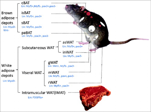
Table 1. Potential myokines, their secretion and target sites, and functions