Figures & data
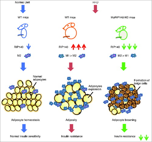
Figure 1. The function of ATM recruitment and contribution to adipose tissue. Left panel: In lean mice fed a normal diet (or ND), the function of ATMs as M2 type maintains adipose tissue homeostasis and normal insulin sensitivity. Middle panel: In HFD-induced obese mice, RIP140 is overexpressed in monocytes/macrophages compared with ND-fed WT mice, resulting in M1 over M2 ATM infiltration and in situ polarization, triggering WAT adiposity and insulin resistance. Right panel: Attenuation of RIP140 in the monocytes/macrophages could result in M2 over M1 ATM polarization, triggering WAT browning and alleviating HFD–induced insulin resistance.