Figures & data
Figure 1. Effect of age, diet and CIT on the expression of UCP1 gene and protein. RET or EPI WAT explants from 2 months old CD (white histogram), 4 months old CD (light gray histogram), 4 months old HFD (dark gray histogram) and 25 months old CD (black histogram) rats were incubated either in the absence of CIT (panels A and B) or with or without CIT (panels C and D) for 8h or 24h as described in materials and methods. Uncoupling protein 1 (UCP1) mRNA levels were evaluated by RT-qPCR. Results were normalized to RPL13 mRNA and expressed in % of control (panels A and C). UCP1 protein levels were estimated by western blots and results were normalized to β-actin (panels B and D). Results represent the mean ± SEM of independent experiments (n = 7) carried out in triplicate from different explants.*P < 0.05 vs. Control; ** P < 0.01 vs. Control. (A) Effects of diet or age on UCP1 gene expression. Histograms represent the ratio of the values obtained from 4-month-old CD rats, 4-month-old HFD rats or 25-month-old rats to the values obtained from 2-month-old CD rats, taken as controls. (B) Effects of diet or age on the expression of UCP1 protein. Histograms represent the relative UCP1 levels to ß-actin. A representative autoradiogram of protein gel blots of UCP1 and β-actin, is shown. (C) Effects of CIT on UCP1 mRNA levels. Histograms represent the ratio of the values obtained from CIT-treated explants from 2-month-old CD rats, 4-month-old CD rats, 4-month-old HFD rats or 25-month-old rats to the values obtained in untreated explants from rats of the corresponding age, taken as controls. (D) Effects of CIT on the expression of UCP1 protein. Histograms represent the relative UCP1 levels to ß-actin. A representative autoradiogram of western blots of UCP1 and β-actin, is shown.
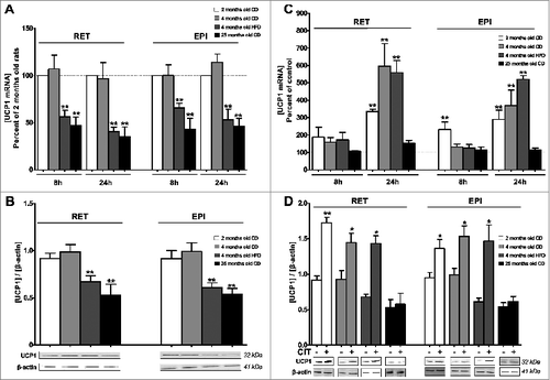
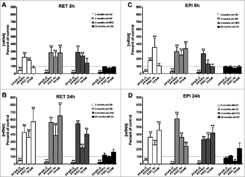
Figure 2. Effect of CIT on the expression of genes coding for transcription factors and regulators involved in mitochondria metabolism. RET (panels A and B) or EPI (panels C and D) WAT explants from 2-month-old CD (white histogram), 4-month-old CD (light gray histogram), 4-month-old HFD (dark gray histogram) and 25-month-old CD (black histogram) rats were incubated with or without CIT for 8h (panels A and C) or 24h (panels B and D), as described in materials and methods. Peroxisome proliferator-activated receptor gamma2 (PPARγ2), peroxisome proliferator-activated receptor α (PPARα), peroxisome proliferator-activated receptor gamma coactivator 1α (PGC-1 α) and mitochondrial transcription factor A (TFAM) mRNA levels were evaluated by RT-qPCR. Histograms represent the ratio of the values obtained from CIT-treated explants from 2-month-old CD rats, 4-month-old CD rats, 4-month-old HFD rats or 25-month-old rats to the values obtained in untreated explants from rats of the corresponding age, taken as controls. Results represent the mean ± SEM of independent experiments (n = 7) carried out in triplicate from different explants, are normalized to RPL13 mRNA and expressed in % of control.*P < 0.05 vs. Control; ** P < 0.01 vs. Control