Figures & data
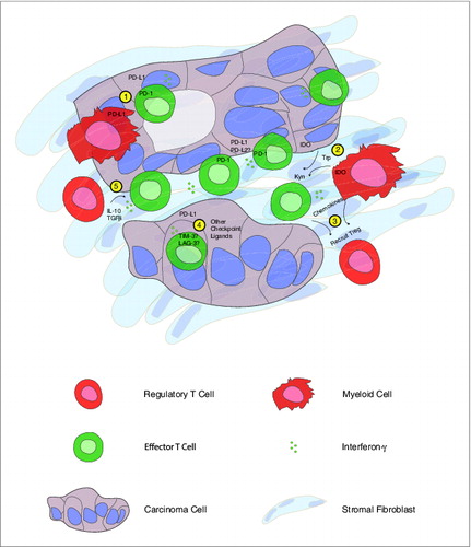
Figure 1. Human pancreatic cancer contains a complex immune infiltrate with both effector and regulatory elements. Based upon our findings, we hypothesize that carcinoma-specific effector CD8+ T cells present throughout the tumor produce interferon γ (IFNγ) that drives immunosuppressive counter-regulatory mechanisms by regulatory T cells (Tregs), myeloid cells, and carcinoma cells. Potential mechanisms of immunosuppression that may serve as therapeutic targets include: 1) inhibition of T-cell activation by stimulation of the negative checkpoint regulatory molecule programmed cell death 1 (PD-1) via binding to PD-1 ligands (PD-L1, PD-L2) expressed on cancer cells or myeloid cells; 2) malignant cell and myeloid cell production of the enzyme indoleamine 2,3-dioxygenase (IDO), which catalyzes the breakdown of tryptophan (Trp) to kynurenine (Kyn); 3) chemokine-mediated recruitment of regulatory T cells; 4) binding of additional T cell co-inhibitory receptors, such as T cell immunoglobulin mucin-3 (TIM-3), lymphocyte activation gene-3 (LAG-3) to ligands displayed by carcinoma cells; 5) production of IL-10 and transforming growth factor β (TGFβ) by Tregs.