Figures & data
Figure 1. Serum concentrations of anti-p21 (CDKN1A) autoantibodies (aAbs) are higher in CRC patients correlating with advanced disease state. (A) Ninety-six well plates were coated with 1 μg/mL of recombinant p21 antigenic peptide (ab56278, Abcam). After blocking and washing steps, plates were incubated with serial dilutions of serum samples from 25 colorectal cancer (CRC) patients vs. serum from 13 normal controls. Binding was detected using horseradish peroxidase labeled goat anti-human Fc antibody and developed per standard protocol. Plates were read on a spectrophotometer at OD450. The optimal serum dilution to obtain 50% binding (OD50) was determined by averaging the results from 3 independent experiments. Statistical analysis was performed by ANOVA. (B-C) Distribution of CRC patients with high and low serum anti-p21 aAb titers, with a threshold half maximal effective concentration (EC50) = 4000, according to the disease stage (B); early, Stages I & II; advanced, Stages III & IV, and according to lymph node (LN) involvement (C). Statistical analysis was performed by 2-sided Fisher's exact test.
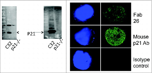
Figure 2. Colorectal cancer patient-derived Fab 26 detects p21 via western blot and Immunofluorescence assays. Colorectal cancer (CRC) patients with high antibody titers were used to derive anti-p21 Fab antibody fragments. Total RNA was isolated from freshly prepared peripheral mononuclear cells, reverse transcribed and subject to PCR to amplify variable heavy chain and light chain antibody regions. (2A) Western blot of cell lysates from C32 CRC cells overexpressing p21 and HCT116 p21−/−, a p21 null CRC cell line, probed by Fab 26 (Left panel) and mouse p21 (Right panel) revealing reliable and specific detection of p21 by Fab 26. (2B) Immunofluorescence staining of HCT116 cells using Fab 26 (upper panel) or monoclonal mouse p21 Ab (middle panel), compared to isotype control in the absence of Fab 26 and presence of secondary and fluorescence Ab (lower panel). Images were taken using confocal microscopy and the 40X objective. The DAPI staining of the stained nuclei are shown on the left for each respective immunofluorescence experiment. The depicted results are representative of 2 independent experiments.