Figures & data
Figure 1. Sarcoidoisis-like lesions occurring in melanoma cancer patient following ipilumumab therapy. Horizontal sections of the brain with melanoma metastatic tumor imaged by magnetic resonance imaging (MRI) and horizontal sections of thorax and abdomen by computed tomography (CT); red arrows indicate isolated sarcoidosis-like granulomas; yellow arrows indicate metastases in the right lung and mediastinum; green arrows indicate residual metastatic burden. Longitudinal imaging: (A) November 2010, pre-ipilimumab; (B) March 2011, after 4 courses of ipilimumab; (C) April 2011, 6 weeks after Ipilimumab; (D) August 2011, 5 months after ipilimumab; (E) November 2012, 20 months after ipilimumab and (F) December 2013, 2 y and 10 months after ipilimumab.
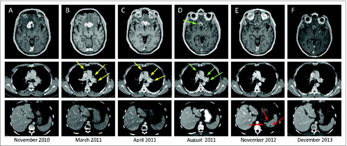
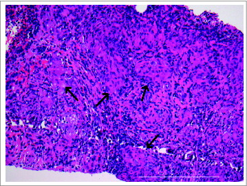
Figure 2. Development of splenic sarcoidosis-like lesions in melanoma patient with durable response to ipilimumab. Ultrasound guided biopsy and histological examination of the hypodense lesions in the spleen of ipilimumab treated melanoma patient showing several non-caseating epithelioid cell granulomas (black arrows) consistent with sarcoidosis (hematoxylin-eosin stain; original magnification × 20, scale bar 500 μm).