Figures & data
Figure 1. Hydrodynamic focusing in a microchannel. Confocal fluorescence microscope image at the junction in MR1 showing the laminar flow of the urea stream by the diluting buffer streams.
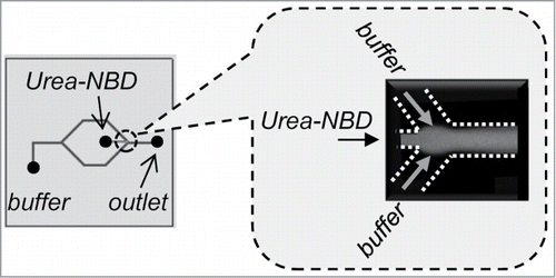
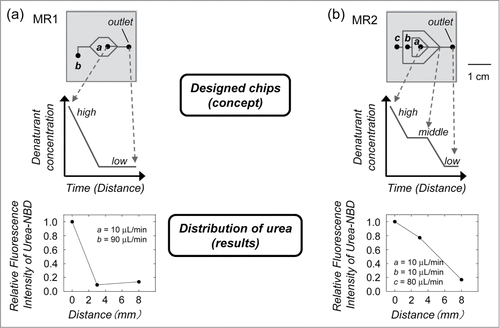
Figure 2. Designed microfluidic chip used for protein refolding. (a) In MR1, the denaturant concentration around the protein rapidly decreases because of diffusion, which is expected to have a similar mechanism to one-step dialysis or dilution. (b) In MR2, the denaturant concentration shows a step-wise decrease, which is similar to step-wise dialysis. The denatured protein was injected into channel a. The dilution buffer was injected into channels b and c. The distributions of denaturant concentrations were measured by the relative fluorescence intensities of the fluorophore in the urea stream as a function of the distance from the inlet.