2,752
Views
66
CrossRef citations to date
0
Altmetric
Review

Peri-adipocyte ECM remodeling in obesity and adipose tissue fibrosis

Pages 89-95 | Published online: 01 Apr 2012

Figures & data

Figure 1. Peri-adipocyte collagens. Left: the scanning electron micrograph of mouse inguinal fat pads; the group of round adipocytes are surrounded with collagen bundles. Middle: immunofluorescent staining of type I collagen (red); thick bundles of type I collagen surrounding the group of adipocytes as well as thinner fibers of type I collagen enwrapping individual adipocyte is displayed. Right: immunofluorescent staining of type IV collagen (green); type IV collagen is found as a component of basement membrane that enwraps each adipocytes; type IV collagen can also be found as the basement membrane underneath the layer of vascular endothelial cells.
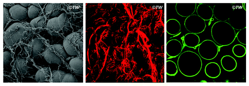
Figure 1. Peri-adipocyte collagens. Left: the scanning electron micrograph of mouse inguinal fat pads; the group of round adipocytes are surrounded with collagen bundles. Middle: immunofluorescent staining of type I collagen (red); thick bundles of type I collagen surrounding the group of adipocytes as well as thinner fibers of type I collagen enwrapping individual adipocyte is displayed. Right: immunofluorescent staining of type IV collagen (green); type IV collagen is found as a component of basement membrane that enwraps each adipocytes; type IV collagen can also be found as the basement membrane underneath the layer of vascular endothelial cells.

Figure 2. Peri-adipocyte ECM proteins. Each adipocyte is surrounded by basement membrane whose framework is defined by type IV collagen. The most abundant fibrillar structure is provided by the cross-linking of triple-helical type I collagen molecules (enlarged in the inset). Types V and VI collagens form micro-fibers that exist between type I collagen fibers as well as between cell surface and type I collagen fibers. Fibronectin is the key ECM protein that defines cell shape and contractility in close association with type I collagen. Matricelluar proteins, i.e., thrombospondins and SPARC, regulate the fibrilliogenesis of type I collagen and interact with multiple ECM proteins.
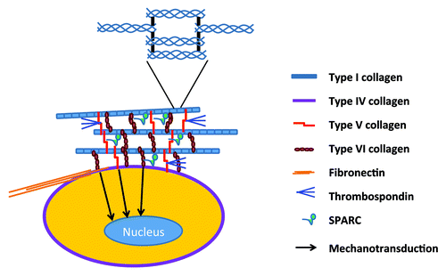
Figure 2. Peri-adipocyte ECM proteins. Each adipocyte is surrounded by basement membrane whose framework is defined by type IV collagen. The most abundant fibrillar structure is provided by the cross-linking of triple-helical type I collagen molecules (enlarged in the inset). Types V and VI collagens form micro-fibers that exist between type I collagen fibers as well as between cell surface and type I collagen fibers. Fibronectin is the key ECM protein that defines cell shape and contractility in close association with type I collagen. Matricelluar proteins, i.e., thrombospondins and SPARC, regulate the fibrilliogenesis of type I collagen and interact with multiple ECM proteins.

Figure 3. MMP14 (MT1-MMP) as a 3D factor. MMP14 is required for the cytoskeletal rearrangement of preadipocytes in a 3D collagen environment (3D preadipocytes). MMP14-dependent cell shape regulation is closely linked to the adipogenic potential of preadipocytes in a 3D environment (3D adipocytes). The loss of MMP14 activity leads to the aberrant cell shape of preadipocytes and impaired 3D adipogenesis, which is coupled with the excess accumulation of undigested collagen fibers. MMP14 is not required for adipogenesis under 2D culture conditions (2D adipogenesis).
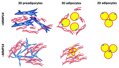
Figure 3. MMP14 (MT1-MMP) as a 3D factor. MMP14 is required for the cytoskeletal rearrangement of preadipocytes in a 3D collagen environment (3D preadipocytes). MMP14-dependent cell shape regulation is closely linked to the adipogenic potential of preadipocytes in a 3D environment (3D adipocytes). The loss of MMP14 activity leads to the aberrant cell shape of preadipocytes and impaired 3D adipogenesis, which is coupled with the excess accumulation of undigested collagen fibers. MMP14 is not required for adipogenesis under 2D culture conditions (2D adipogenesis).

Figure 4. Nutritional injury and genetics in adipose tissue pathology. Nutritional injury, such as high-fat diet, unravels genetic predispositions of individuals. Adipose tissues can maintain healthy tissue function through physiological tissue regeneration in response to nutritional injury; however, in certain individuals, adipose tissues are genetically predisposed to fibrosis (overhealing) or inflammation (chronic wound).
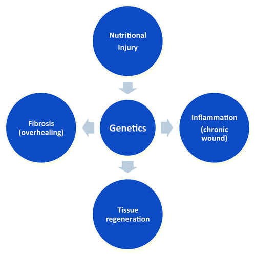
Figure 4. Nutritional injury and genetics in adipose tissue pathology. Nutritional injury, such as high-fat diet, unravels genetic predispositions of individuals. Adipose tissues can maintain healthy tissue function through physiological tissue regeneration in response to nutritional injury; however, in certain individuals, adipose tissues are genetically predisposed to fibrosis (overhealing) or inflammation (chronic wound).