Figures & data
Figure 1. Schematic model showing the AdipoChaser mouse model and adipogenesis found in different fat depots. (A) The AdipoChaser mouse model: Prior to doxycycline treatment, every white adipocyte is LacZ-negative (adipocytes surrounded by red circles). After doxycycline exposure, all white adipocytes are LacZ-positive (adipocytes surrounded by blue circles). After doxycycline withdrawal, if mice are kept under conditions that induce adipogenesis, new adipocytes will be observed as LacZ-negative adipocytes (adipocytes surrounded by red circles with red glow). (B) Adipogenesis in eWAT in adult mouse: Prolonged HFD feeding (more than 1 mo), cold exposure, and β-3 treatment induce adipogenesis (white adipocyte) in eWAT. (C) Adipogenesis in sWAT in the adult mouse: HFD feeding only induces hypertrophy in sWAT; cold exposure and β-3 treatment-induced beige adipocytes within sWAT are from de novo adipogenesis. (D) The development of eWAT: Adipocytes in the eWAT are differentiated postnatally between birth and sexual maturation. LacZ expression in adipocytes is pulsed at postnatal day (P) 10 (doxycycline withdrawal), new white adipocytes are observed at P28 and there are more white adipocytes by P56. (E) The development of sWAT: All the adipocytes in the sWAT start to differentiate between embryonic day (E) 14 and E18, but the differentiation takes much longer and finishes postnatally. LacZ expression in adipocytes is pulsed at E18 (doxycycline withdrawal), no new white adipocytes are observed at P28 or P56.
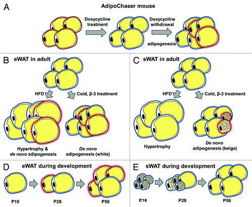
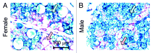
Figure 2. Newly differentiated adipocytes are not necessarily small in size. (A and B) Representative β-gal staining of sWAT from 28 d old female and male mice. The mothers of these mice were on doxycycline diet during embryonic day 9 to embryonic day 16. Pink arrows, LacZ-negative adipocytes that have relative large cell sizes; blue arrows, LacZ-positive adipocytes that have relative small cell sizes.