1,063
Views
47
CrossRef citations to date
0
Altmetric
Autophagic Punctum

Processing of proteins in autophagy vesicles of antigen-presenting cells generates citrullinated peptides recognized by the immune system

&
Pages 429-430 | Published online: 24 Feb 2012

Figures & data

Figure 1. Model of citrullination by (A) DC and macrophages, or (B) B cells. (A and B) represent the possible scenarios taking place in APC that result in citrullination of peptide-MHC complexes (pMHC). (A) represents the pathways in DC or macrophages. These cells undergo constitutive autophagy and present the citrullinated peptides after the protein antigen comes into contact with the autophagy vesicles (AV). In (A), the protein antigen lysozyme (HEL) binds to the plasma membrane, and traffics to early endosomal vesicles (EV). From the EV it follows two pathways. In one it is taken to late vesicles (LV), processed to peptides and presented at the cell surface as a pMHC complex. In the second pathway, the protein is taken to an autophagic vesicle (AV) where it is processed to peptides, some of which are citrullinated and presented at the cell surface (cit-pMHC). (B) represents the pathways in B cells. Citrullination in B cells occurs only after autophagy is induced by engagement by antigen of the B cell receptor, shown as an immunoglobulin (Ig) molecule on the cell membrane. Receptor engagement leads to striking subcellular changes that lead to colocalization of AV and antigen-processing compartments, and presentation of citrullinated peptides. (B) shows that the antigen HEL can enter the B cell directly after interaction with plasma membrane, and is taken to EV and LV, to generate unmodified pMHC complexes. However, when HEL is bound to the surface Ig molecules, autophagy is induced, and some of the HEL-Ig complex then traffics to the AV to be processed, and generates the cit-pMHC complex.
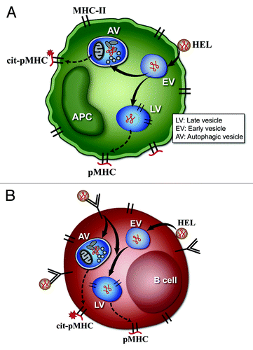
Figure 1. Model of citrullination by (A) DC and macrophages, or (B) B cells. (A and B) represent the possible scenarios taking place in APC that result in citrullination of peptide-MHC complexes (pMHC). (A) represents the pathways in DC or macrophages. These cells undergo constitutive autophagy and present the citrullinated peptides after the protein antigen comes into contact with the autophagy vesicles (AV). In (A), the protein antigen lysozyme (HEL) binds to the plasma membrane, and traffics to early endosomal vesicles (EV). From the EV it follows two pathways. In one it is taken to late vesicles (LV), processed to peptides and presented at the cell surface as a pMHC complex. In the second pathway, the protein is taken to an autophagic vesicle (AV) where it is processed to peptides, some of which are citrullinated and presented at the cell surface (cit-pMHC). (B) represents the pathways in B cells. Citrullination in B cells occurs only after autophagy is induced by engagement by antigen of the B cell receptor, shown as an immunoglobulin (Ig) molecule on the cell membrane. Receptor engagement leads to striking subcellular changes that lead to colocalization of AV and antigen-processing compartments, and presentation of citrullinated peptides. (B) shows that the antigen HEL can enter the B cell directly after interaction with plasma membrane, and is taken to EV and LV, to generate unmodified pMHC complexes. However, when HEL is bound to the surface Ig molecules, autophagy is induced, and some of the HEL-Ig complex then traffics to the AV to be processed, and generates the cit-pMHC complex.

Reprints and Corporate Permissions

Please note: Selecting permissions does not provide access to the full text of the article, please see our help page How do I view content?

To request a reprint or corporate permissions for this article, please click on the relevant link below:

Academic Permissions

Please note: Selecting permissions does not provide access to the full text of the article, please see our help page How do I view content?

Obtain permissions instantly via Rightslink by clicking on the button below:

If you are unable to obtain permissions via Rightslink, please complete and submit this Permissions form. For more information, please visit our Permissions help page.