Figures & data
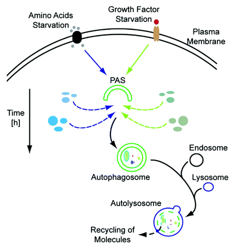
Figure 1. Autophagosomal protein dynamics. Depending on the inducing stimulus, e.g., amino acid deprivation (left side) or growth factor starvation (right side), the autophagosomal protein composition may vary. Degradation of cellular material which is not needed during the respective condition, exemplified by blue and green ellipses for amino acid and growth factor starvation, respectively, will be preferred. In addition, the time frame of stimulation is also reflected by the autophagosomal proteome. PAS, phagophore assembly site.