Figures & data
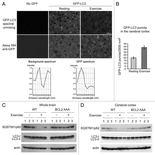
Figure 1. Autophagy is induced by exercise in the brain. (A) Representative images of GFP-LC3 puncta in the cerebral cortex from wild-type GFP-LC3 mice at rest and after 95 min of exercise. The top panel shows the GFP-LC3 puncta in the brain after the auto-fluorescent signals were removed by spectral unmixing analyses. For spectral unmixing, images were acquired under a Zeiss 63x 1.4 NA PLAN-APOCHROMAT objective using a Zeiss LSM510 microscope equipped with a META multispectral detector at excitation of 488 nm. Linear unmixing was performed using the Zeiss LSM software by subtracting background (no GFP) peaks of 533 nm and 575 nm from the GFP spectrum, which peaked at 520 nm in tissue sections from GFP-LC3 mice. The bottom panel shows representative images of GFP immunofluorescence staining in cerebral crotex from the same wild-type GFP-LC3 at rest and after 95 min of exercise. Brain sections were stained with an anti-GFP antibody and an Alexa Fluor 594-conjugated secondary antibody. Scale bar: 20 μm. (B) Quantification of GFP-LC3 puncta shown in (A). A minimum of 8 fields per mouse was analyzed. Results shown represent mean ± s.d. for 3 mice per experimental group. (C and D) Biochemical analysis of autophagy induction in extracts of whole brain (C) or cerebral cortex (D) from wild-type (WT) and BCL2 AAA mice at rest (-) or after 95 min of exercise by western blot detection of SQSTM1/p62 and LC3-II conversion. Actin was used as a loading control. Three mice per experimental group are shown.