Figures & data
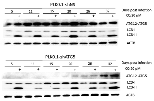
Figure 1. LC3-II formation is blocked only temporarily after ATG5 knockdown. Western blot analysis is shown of HeLa cells treated for 6 h ± 20 µM chloroquine to block basal autophagic flux of PLK0.1-shRNA nonsilencing (control shRNA) compared with shRNA targeting ATG5 cultured postinfection for up to 32 d. Although the knockdown worked well at 5 d postinfection through 26 d, basal autophagy was only blocked effectively in the samples tested at 11 and 15 d after infection.