4,131
Views
36
CrossRef citations to date
0
Altmetric
Views and Commentaries

Evolutionarily conserved role and physiological relevance of a STX17/Syx17 (syntaxin 17)-containing SNARE complex in autophagosome fusion with endosomes and lysosomes

, , &
Pages 1642-1646 | Received 17 May 2013, Accepted 09 Jul 2013, Published online: 22 Jul 2013

Figures & data

Figure 1. Ultrastructural images of wild-type and Syx17 mutant Drosophila cells from early L3 stage larvae starved for 3 h. (A) Both autophagosomes (AP) and electron-dense autolysosomes (AL) can be observed in various tissues of starved control larvae, such as the fat body shown here. M, mitochondrion. (B) Typical double-membrane autophagosomes accumulate in large numbers in starved Syx17 mutant larvae, illustrated by a Malpighian tubule cell in this image. (C) This panel shows a rarely observed phagophore in a Syx17 mutant cell, which appears as a curved, nonbranching membrane cistern with a characteristic cleft between the two membranes after standard glutaraldehyde fixation. While the empty-looking space is an artifact, it greatly facilitates the identification of early autophagic structures. Arrowheads point to the edges of the phagophore, where the transitions of prospective outer and inner membrane layers are clearly visible.
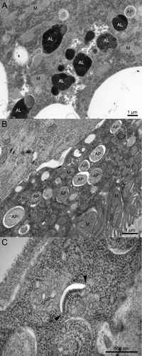
Figure 1. Ultrastructural images of wild-type and Syx17 mutant Drosophila cells from early L3 stage larvae starved for 3 h. (A) Both autophagosomes (AP) and electron-dense autolysosomes (AL) can be observed in various tissues of starved control larvae, such as the fat body shown here. M, mitochondrion. (B) Typical double-membrane autophagosomes accumulate in large numbers in starved Syx17 mutant larvae, illustrated by a Malpighian tubule cell in this image. (C) This panel shows a rarely observed phagophore in a Syx17 mutant cell, which appears as a curved, nonbranching membrane cistern with a characteristic cleft between the two membranes after standard glutaraldehyde fixation. While the empty-looking space is an artifact, it greatly facilitates the identification of early autophagic structures. Arrowheads point to the edges of the phagophore, where the transitions of prospective outer and inner membrane layers are clearly visible.