Figures & data
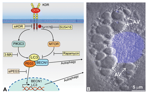
Figure 1. Soluble DCN evokes endothelial cell autophagy. (A) Schematic representation showing how high-affinity interactions between DCN and KDR ectodomain (Ig3–5) trigger a signal cascade simultaneously activating and suppressing PIK3C3 and MTOR, respectively. This DCN-evoked process is blocked by siRNA targeting KDR (siKDR) or by KDR tyrosine kinase-inhibitor SU5416. The rapidly-induced PEG3 physically associates with BECN1 and LC3 for autophagic induction. Moreover, unbound PEG3 presumably undergoes nuclear translocation to transactivate key genetic loci including BECN1 and LC3, and this process is inhibited by siRNA targeting PEG3 (siPEG3). This reinforced network, with PEG3 serving as the primary node, permits DCN to evoke protracted endothelial cell autophagy as the underlying basis for angiostasis. (B) Differential interference contrast microscopy of a mouse microvascular endothelial cell following an 18 h treatment with DCN (200 nM) in nutrient-rich conditions. The nucleus is stained in blue by DAPI. Notice the formation of multiple cytoplasmic autophagic vacuoles (AV).