2,032
Views
13
CrossRef citations to date
0
Altmetric
Protocol

Detection of Saccharomyces cerevisiae Atg13 by western blot

, , &
Pages 514-517 | Received 20 Nov 2013, Accepted 02 Jan 2014, Published online: 09 Jan 2014

Figures & data

Figure 1. Detection of overexpressed Atg13 using rabbit polyclonal antiserum. Yeast cells overexpressing Atg13 were grown in SD and then shifted to SD-N at time 0. Samples were collected at the indicated time points and analyzed by SDS-PAGE and western blot using antiserum to Atg13. This figure is a modification of data previously published in reference Citation8, and is reproduced by permission of the American Society for Biochemistry and Molecular Biology and Elsevier, copyright 2000.
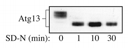
Figure 1. Detection of overexpressed Atg13 using rabbit polyclonal antiserum. Yeast cells overexpressing Atg13 were grown in SD and then shifted to SD-N at time 0. Samples were collected at the indicated time points and analyzed by SDS-PAGE and western blot using antiserum to Atg13. This figure is a modification of data previously published in reference Citation8, and is reproduced by permission of the American Society for Biochemistry and Molecular Biology and Elsevier, copyright 2000.

Figure 2. Wild-type yeast cells expressing Atg13 under the endogenous promoter from a multicopy plasmid (YEp351[APG13]) were grown in SD for 8 h and treated with rapamycin for 15 min as indicated. Protein extracts were analyzed by western blot using anti-Atg13 serum.

Figure 2. Wild-type yeast cells expressing Atg13 under the endogenous promoter from a multicopy plasmid (YEp351[APG13]) were grown in SD for 8 h and treated with rapamycin for 15 min as indicated. Protein extracts were analyzed by western blot using anti-Atg13 serum.

Figure 3. Comparison between different methods to detect Atg13. Wild-type cells (BY4741) were transformed with a vector expressing HA-Atg13 under its own promoter (pHC078). Cultures were grown until mid-log phase in SD and starved for nitrogen 2 h (SD-N). Lanes 1 and 2 correspond to cells that were lysed with NP-40 buffer and disrupted with glass beads; cells were harvested by centrifugation, resuspended in NP-40 lysis buffer (200 µl) containing protease and phosphatase inhibitors, followed by glass bead lysis and centrifugation at 700 × g for 3 min. Proteins were quantified by the BCA assay. The same amount of protein was loaded onto the gel. Lanes 3 and 4 correspond to cells that were disrupted by vortex with glass beads and protein sample buffer (PSB). Lanes 5 to 8 samples were prepared using the TCA-MURB method described in this paper. The protein extracts of lane 7 and 8 were mixed with PSB.
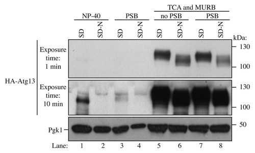
Figure 3. Comparison between different methods to detect Atg13. Wild-type cells (BY4741) were transformed with a vector expressing HA-Atg13 under its own promoter (pHC078). Cultures were grown until mid-log phase in SD and starved for nitrogen 2 h (SD-N). Lanes 1 and 2 correspond to cells that were lysed with NP-40 buffer and disrupted with glass beads; cells were harvested by centrifugation, resuspended in NP-40 lysis buffer (200 µl) containing protease and phosphatase inhibitors, followed by glass bead lysis and centrifugation at 700 × g for 3 min. Proteins were quantified by the BCA assay. The same amount of protein was loaded onto the gel. Lanes 3 and 4 correspond to cells that were disrupted by vortex with glass beads and protein sample buffer (PSB). Lanes 5 to 8 samples were prepared using the TCA-MURB method described in this paper. The protein extracts of lane 7 and 8 were mixed with PSB.

Reprints and Corporate Permissions

Please note: Selecting permissions does not provide access to the full text of the article, please see our help page How do I view content?

To request a reprint or corporate permissions for this article, please click on the relevant link below:

Academic Permissions

Please note: Selecting permissions does not provide access to the full text of the article, please see our help page How do I view content?

Obtain permissions instantly via Rightslink by clicking on the button below:

If you are unable to obtain permissions via Rightslink, please complete and submit this Permissions form. For more information, please visit our Permissions help page.