Figures & data
Figure 1. Novel nuclear envelope localization of IQGAP1. (A) Nuclear halo staining of IQGAP1 occurs in several cell lines. Deconvolution fluorescence cell images of human epithelial-derived MCF-7 and HT29 cancer cells and mouse fibroblast NIH 3T3 cells. Cells were immunolabelled and stained for IQGAP1 (H-109, Santa-Cruz; red), F-actin (phalloidin-FITC; green) and DNA (Hoechst; blue). (B) IQGAP1 locates to the cytoplasmic face of the outer nuclear membrane. Deconvolution microscopy fluorescence cell images of MCF-7 cells stained with IQGAP1 (red) and FxFG-repeat nucleoporins (mAb414; green). The graph depicts pixel intensity of IQGAP1 (red) and nucleoporin (green) staining from the indicated cross-section in the enlarged micrograph (toward cytoplasm on right). (C) High resolution scoring of IQGAP1 co-localization with nuclear envelope in MCF-7 cells (n = 62). (D) Electron micrographs of ultrathin cryosections of MCF-7 cells immunolabelled with IQGAP1 pAb. Cells with no primary antibody showed no specific staining pattern (not shown). Thin closed arrow indicates nuclear rim; broad open arrow indicates immunogold-labeling of IQGAP1. White bar, 200 nm.
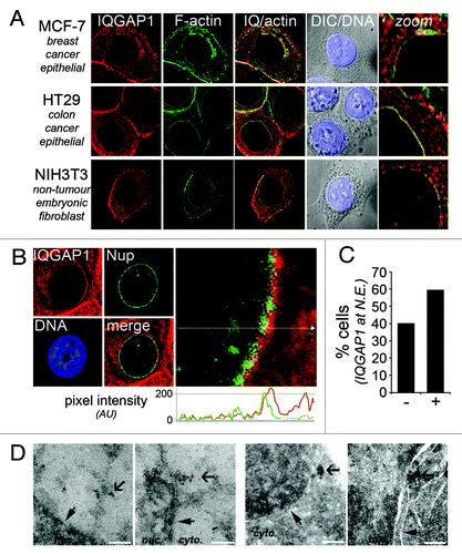
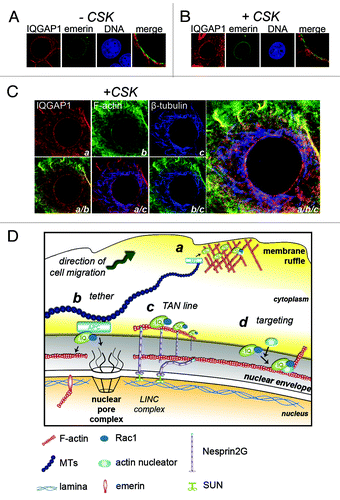
Figure 2. IQGAP1 is associated with cytoskeletal architecture at the nuclear envelope. (A and B) IQGAP1 is anchored at the nuclear rim. MCF-7 cells transiently expressing emerin-GFP were either (A) fixed-first (-CSK) and detergent permeabilized or (B) CSK-extracted (+CSK) and fixed, then stained for IQGAP1 (red). (C) Confocal fluorescence microscopy images of CSK-extracted MCF-7 cells tri-labeled for IQGAP1 (a; red), F-actin (b; green) and β-tubulin (c; blue). Bottom panel and enlarged image on right show merged fluorescence micrographs. (D) Schematic proposing the roles of IQGAP1 at the cytoplasmic face of the nuclear rim. a, At plasma membrane sites involved in cell migration IQGAP1 tethers microtubule networks to the cortical actin mesh via the MT +end protein APC for cell polarization cues. b, Tethering for polarization cues? At the cytoplasmic face of the NE, IQGAP1 overlays with MTs and actin and may tether these cytoskeletal networks via APC. c, Nuclear repositioning? TAN lines in mesenchymal cells assist in nuclear repositioning during cell polarization and cell migration. d, Orchestrating actin polymerisation and rearrangements? IQGAP1 targets numerous actin-associated proteins to subcellular sites. IQGAP1 anchors actin-branching and nucleating proteins to membrane ruffles and to yeast cytokinetic ringsCitation9 to orchestrate actin rearrangements. IQGAP1 may target other proteins to the nuclear envelope during cell migration or cell cycle events such as during nuclear envelope breakdown.