Figures & data
Figure 1. Filopodia from COS7 cells co-expressing GFP-Myo3a and cherry-Myo3b consistently display GFP-Myo3a accumulated at their extreme tips, while cherry-Myo3b consistently trails behind Myo3a with a relatively longer tip-to-base decay length (left column). GFP-Myo3a with and without the 3THDII actin-binding site (GFP-Myo3aΔTHDII) consistently accumulates at filopodia tips ahead of GFP-Myo3b (left and middle columns, respectively), while GFP-Myo3a does not exclude GFP-Myo3aΔTHDII (right column). The black and gray boxes are the motor domains of Myo3a and Myo3b, respectively. The red crescents represent the IQ domains.
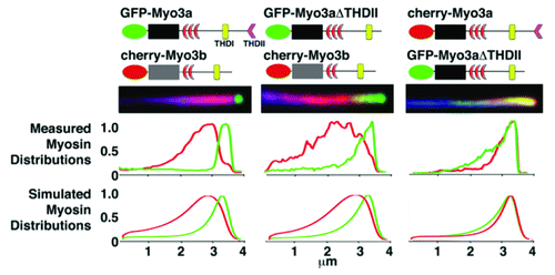
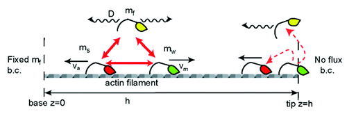
Figure 2. Schematic description of the calculated model of myosin distributions. We consider freely diffusing (yellow), processively walking (green) and stalled (red) myosins, that convert into each other (red arrows) with defined reaction coefficients. At the protrusion tip we illustrate the two types of boundary conditions that we used; walking motors detach and diffuse freely from the tip, or become stalled and are carried away by the actin treadmilling.