Figures & data
Figure 1. Viability of soft tissue sarcoma STSA-1 (A), melanoma CHAS (B), osteosarcoma D17 (C) and prostate carcinoma DT08/40 (D). cells after LIVP6.1.1 infection at MOIs of 1.0 and 0.1. Viable cells after infections with LIVP6.1.1virus at MOIs of 0.1 and 1.0 were detected using a XTT assay (Cell Proliferation Kit II, Roche Diagnostics, Mannheim, Germany)(Sigma, Taufkirchen, Germany). Mean values (n = 3) and standard deviations are shown as percentages of respective controls. The data represent three independent experiments
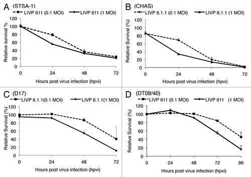
Figure 2. Replication capacity of the vaccinia virus strains LIVP6.1.1 and GLV-1h68 in different canine cancer cells. For the viral replication assay, STSA-1 (A) and DT08/40 (B) cells grown in 24-well plates were infected with either LIVP6.1.1 or GLV-1h68 at an MOI of 0.1. Cells and supernatants were collected for the determination of virus titers at various time points. Viral titers were determined as pfu per ml in triplicates by standard plaque assay in CV-1 cell monolayers. Averages plus standard deviation are plotted. The data represent three independent experiments.
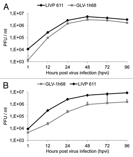
Figure 3. Effects of systemic LIVP6.1.1 virus injection on tumor growth (A, B) and the body weights (C, D) of STSA-1 or DT08/40 xenografted mice. Two groups each of (A, C) STSA-1 tumor-bearing nude mice (n = 6) and (B, D) DT08/40 tumor bearing mice (n = 6) were either treated with a single dose of 5x10⁶ pfu LIVP6.1.1 or with PBS (mock control) intravenously (iv). The statistical significance was confirmed by Student’s t-test where * and ** indicate p < 0.05 and 0.01 respectively.
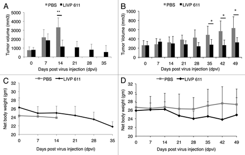
Table 1. Biodistribution of LIVP6.1.1 in virus-treated STSA-1 xenografted mice at 35 d post virus injection (dpvi)
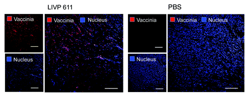
Figure 4. Immunohistochemical staining of infected and uninfected STSA-1 xenograft tumors at 35 dpvi. Tumor-bearing mice were either mock treated (PBS) or infected with LIVP6.1.1. Tumor sections were labeled with anti-vaccinia virus antibodies (red) and nuclei were stained with Hoechst (blue). Scale bars, 2.5 mm.