896
Views
8
CrossRef citations to date
0
Altmetric
Research Paper

Phosphate enhances levan production in the endophytic bacterium Gluconacetobacter diazotrophicus Pal5

, , &
Pages 173-179 | Received 03 Mar 2014, Accepted 04 Apr 2014, Published online: 09 Apr 2014

Figures & data

Table 1. Composition of LGI-P, Y&P-NaCl, high P, and low P media

Figure 1.G. diazotrophicus Pal5 WT, gumD mutant, and lsdA mutant strains grown on high or low P solid medium. Plus (+++) indicates the strongest mucous trait, plus (+) is moderate, and minus (-) is the weakest. Cells were grown for 1 wk at 30 °C.
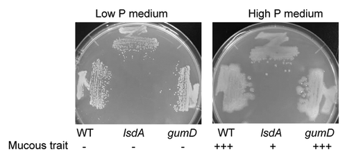
Figure 1.G. diazotrophicus Pal5 WT, gumD mutant, and lsdA mutant strains grown on high or low P solid medium. Plus (+++) indicates the strongest mucous trait, plus (+) is moderate, and minus (-) is the weakest. Cells were grown for 1 wk at 30 °C.

Figure 2.G. diazotrophicus Pal 5 WT grown on high P, low P, and low P medium + NaCl containing the same concentration of Na+ as that of the high P medium. Cells were grown for 1 wk at 30 °C.
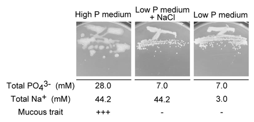
Figure 2.G. diazotrophicus Pal 5 WT grown on high P, low P, and low P medium + NaCl containing the same concentration of Na+ as that of the high P medium. Cells were grown for 1 wk at 30 °C.

Figure 3. HPLC analysis of mucous materials. Authentic levan and mucous material collected from colonies of WT, gumD, and lsdA cells grown for 1 wk at 30 °C on the indicated solid media were hydrolyzed and analyzed. The vertical axis of the chromatograph shows the relative peak level. The retention time (min) is shown on the horizontal axis. Arrows show the levan peak (15.3 min).
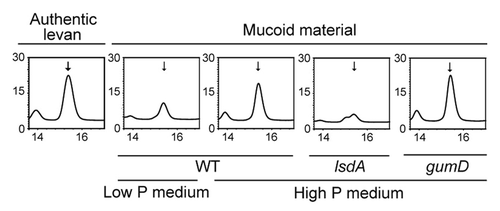
Figure 3. HPLC analysis of mucous materials. Authentic levan and mucous material collected from colonies of WT, gumD, and lsdA cells grown for 1 wk at 30 °C on the indicated solid media were hydrolyzed and analyzed. The vertical axis of the chromatograph shows the relative peak level. The retention time (min) is shown on the horizontal axis. Arrows show the levan peak (15.3 min).

Table 4. Concentration of phosphate in sugarcane juice

Figure 4. Growth on high P or low P medium of bacteria isolated from sugarcane juice. Plus (+++) indicates the strongest mucous trait, plus (+) is moderate, and minus (-) is the weakest. Cells were grown for 1 wk at 30 °C. Bacteria were tentatively identified based on the rDNA sequences.
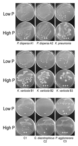
Figure 4. Growth on high P or low P medium of bacteria isolated from sugarcane juice. Plus (+++) indicates the strongest mucous trait, plus (+) is moderate, and minus (-) is the weakest. Cells were grown for 1 wk at 30 °C. Bacteria were tentatively identified based on the rDNA sequences.

Figure 5. Growth of G. diazotrophicus Pal5 WT and lsdA disruptant on solid media containing hydrogen peroxide (H2O2). Three spotted cells were diluted 10-fold from OD600 of 1.0 to 0.01 from right to left.
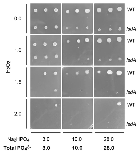
Figure 5. Growth of G. diazotrophicus Pal5 WT and lsdA disruptant on solid media containing hydrogen peroxide (H2O2). Three spotted cells were diluted 10-fold from OD600 of 1.0 to 0.01 from right to left.

Table 2. Primers used in the study a

Table 3. Plasmids used in the study

Supplemental material

Additional material

Download Zip (243.3 KB)

Reprints and Corporate Permissions

Please note: Selecting permissions does not provide access to the full text of the article, please see our help page How do I view content?

To request a reprint or corporate permissions for this article, please click on the relevant link below:

Academic Permissions

Please note: Selecting permissions does not provide access to the full text of the article, please see our help page How do I view content?

Obtain permissions instantly via Rightslink by clicking on the button below:

If you are unable to obtain permissions via Rightslink, please complete and submit this Permissions form. For more information, please visit our Permissions help page.