Figures & data
Figure 1. Affinity-based NGF delivery from PEG-co-peptide CS system. Eight-arm PEG (black lines) are modified with bi-functional cross-linking peptides (dark gray dumbbells) on 6 arms and CS-binding peptides (light gray hexagons) on 2 arms. CS (striped stars) interacts with CS-binding peptides and NGF (spotted triangles).
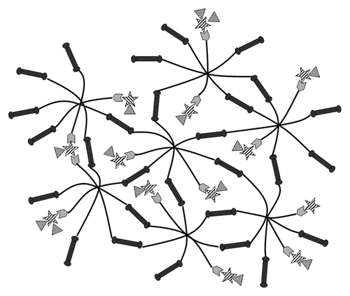
Table 1. Gel compositions
Figure 2. Frequency sweep (0.5–500 rad/s) of gels at 0.5% strain. PEG gels without C6S or BP had the highest viscoelastic properties while gels with C6S and/or BP had significantly lower complex moduli. Mean ± SE.
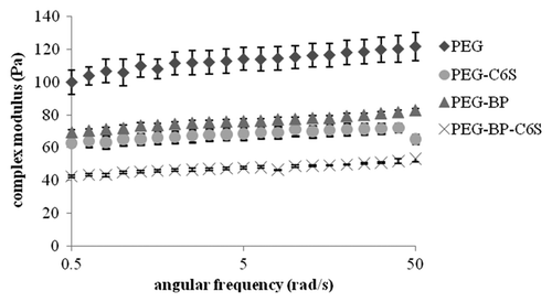
Figure 3. Time sweep of PEG gels at 10 rad/s and 0.5% strain. PEG gels that contained C6S and/or BP had significantly lower complex moduli than gels without C6S or BP. Mean ± SE, *p < 0.05 different relative to PEG.
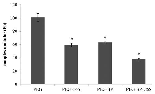
Figure 4. NGF release profile of gels with and without BP and C6S. NGF release was monitored over 48 h. After 2 d, the gels were digested and the amount of NGF quantified. PEG gels that include BP and C6S had the slowest release while PEG gels that only had C6S had the fastest release. Mean ± SE.
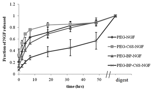
Table 2. Summary of post-hoc test of NGF release from different gels
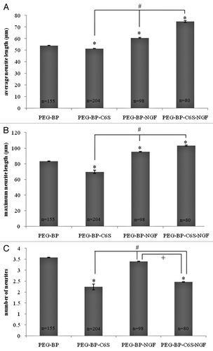
Figure 5. Effect of C6S and NGF on cortical neuron outgrowth. Neurons were cultured on gels with and without C6S and/or NGF for 48 h. The average neurite length (A), maximum neurite length (B), and number of neurites (C) were determined for each gel composition. Mean ± SE, *p < 0.05 relative to PEG-BP, #p < 0.05 relative to PEG-BP-C6S, +p < 0.05 relative to PEG-BP-NGF.