2,805
Views
62
CrossRef citations to date
0
Altmetric
Review

Actin binding proteins

Their ups and downs in metastatic life

Pages 199-213 | Received 27 Sep 2012, Accepted 07 Dec 2012, Published online: 09 Jan 2013

Figures & data

Figure 1. Actin organization in migrating cancer cell. (A) Staining for F-actin using Phalloidin-Alexa488 in a migrating Rama 37 malignant cell expressing high levels of S100A4. In this image, the structures of lamellipodium/lamellum and filopodium are clearly visible at the leading edge of the cell. (B and C) present models for the lamellipodium/lamellum and filopodium and the respective molecular organization within, focusing on the proteins presented in this review. (B) A simplified model for lamellipodium/lamellum formation. In the lamellipodium, free barbed ends of actin filaments recruit the Arp2/3 complex via activation by WASP/WAVE complex and cortactin. The Arp2/3 complex nucleates a new actin filament from the side of existing filaments and remains at the branching point. In the lamellum, actin filaments are bound to tropomyosins, preventing interactions with other actin binding proteins. (C) A simplified model for filopodia formation. Individual filaments of the filopodium emerge from the branching point on other filaments, through actin polymerization promoted by the Arp2/3 complex. Further addition of actin monomers at the barbed end of actin filaments is nucleated by the formin family, whereas fascin regulates filopodia stability through its bundling activities.
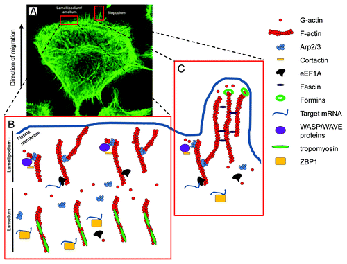
Figure 1. Actin organization in migrating cancer cell. (A) Staining for F-actin using Phalloidin-Alexa488 in a migrating Rama 37 malignant cell expressing high levels of S100A4. In this image, the structures of lamellipodium/lamellum and filopodium are clearly visible at the leading edge of the cell. (B and C) present models for the lamellipodium/lamellum and filopodium and the respective molecular organization within, focusing on the proteins presented in this review. (B) A simplified model for lamellipodium/lamellum formation. In the lamellipodium, free barbed ends of actin filaments recruit the Arp2/3 complex via activation by WASP/WAVE complex and cortactin. The Arp2/3 complex nucleates a new actin filament from the side of existing filaments and remains at the branching point. In the lamellum, actin filaments are bound to tropomyosins, preventing interactions with other actin binding proteins. (C) A simplified model for filopodia formation. Individual filaments of the filopodium emerge from the branching point on other filaments, through actin polymerization promoted by the Arp2/3 complex. Further addition of actin monomers at the barbed end of actin filaments is nucleated by the formin family, whereas fascin regulates filopodia stability through its bundling activities.

Table 1. Regulation of specific actin binding proteins in cancer tissue samples and cell lines

Table 2. miRNAs dependent mechanisms regulating the levels of specific actin binding proteins in different cancer samples and cell lines

Reprints and Corporate Permissions

Please note: Selecting permissions does not provide access to the full text of the article, please see our help page How do I view content?

To request a reprint or corporate permissions for this article, please click on the relevant link below:

Academic Permissions

Please note: Selecting permissions does not provide access to the full text of the article, please see our help page How do I view content?

Obtain permissions instantly via Rightslink by clicking on the button below:

If you are unable to obtain permissions via Rightslink, please complete and submit this Permissions form. For more information, please visit our Permissions help page.