Figures & data
Figure 1. Axial (A–E) fluid attenuation inversion recovery (FLAIR) MRI Brain and sagittal T2-weighted MRI of the cervical cord (1–5) demonstrate a stable distribution of lesions from 2007 (A and 1) to 2012 (F and 6).
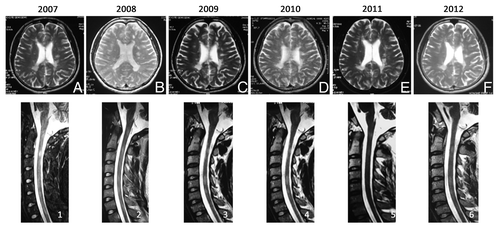
Table 1. Summary of MSC treatment, adverse events and clinical disability score (EDSS)
Supplemental material