Figures & data
Figure 1. Double immunofluorescence staining of fibrillin-1 (green) and α8 integrin (red). A, B, C kidney section from control rat. D, E, F kidney section from rat after 7 d of Thy1.1 nephritis. Colocalization is indicated by yellow color.
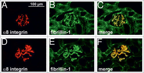
Figure 2. Real-time RT-PCR expression analysis of fibrillin-1 and α8 integrin in the renal cortex of control rats (co) and rats after 7 days of Thy1.1 nephritis (d7) Data are means ± sd. * P < 0.05 vs. respective control.
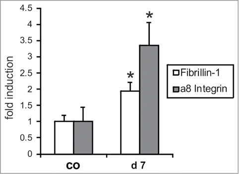
Figure 3. Attachment of wild type (wt) and α8 integrin-deficient (a8−/−) mesangial cells on fibrillin-1 fragment. A: evaluation of cells adhered to fibrillin-1. B: evaluation of cells spread on fibrillin-1. Coating with bovine serum albumin (BSA) served as a negative control, coating with fibronectin (FN) served as a positive control. Results are representative for 3 independent experiments. Data are means ± sd. * P < 0.05 vs. wt.
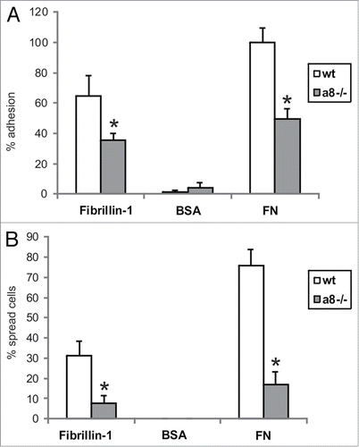
Figure 4. Attachment of wild type (wt) and α8 integrin-deficient (a8−/−) mesangial cells on fibrillin-1 fragment after blocking of αv integrin. Coating with bovine serum albumin (BSA) served as a negative control. Results are representative for 3 similar experiments. Data are means ± sd. * P < 0.05 vs. BSA control, # P < 0.05 vs. wt, § P < 0.05 vs. a8−/− on fibrillin-1.
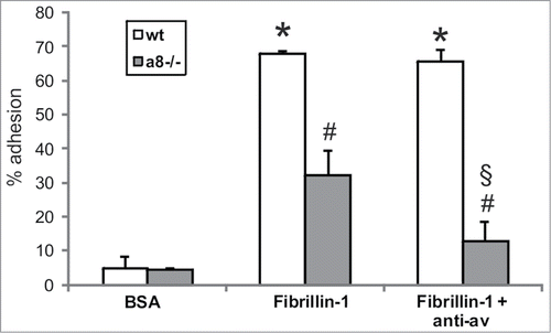
Figure 5. Staining of α8 integrin in wild type mesangial cells attached to fibrillin-1 (A), to fibronectin (α8 integrin ligand) as a positive control (B) to bovine serum albumin (C) as a negative control, and to collagen I (not a ligand for α8 integrin; D).
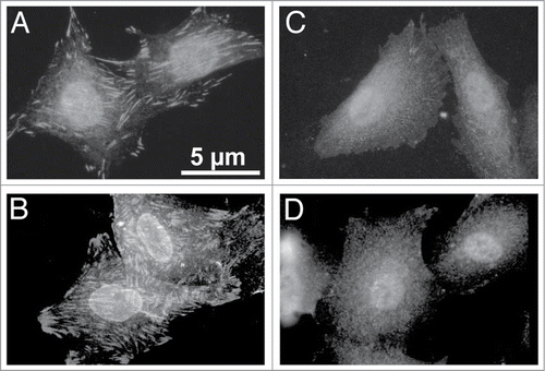
Figure 6. Migration of wild type (wt) and α8 integrin-deficient (a8−/−) mesangial cells on fibrillin-1 fragment (A). B: Migration on BSA as a negative control. C: Migration on fibronectin (FN) as a positive control. Results are representative for 3 similar experiments. Data are means ± sd.
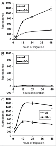
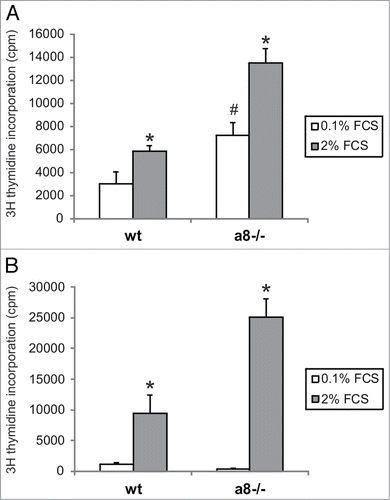
Figure 7. Proliferation of wild type (wt) and α8 integrin-deficient (a8−/−) mesangial cells seeded on fibrillin-1 fragment (A). Cells were serum starved in medium containing 0.1% fetal calf serum (FCS). Proliferation was stimulated with addition of 2% fetal calf serum. B: Proliferation on fibronectin was evaluated as a positive control. Data are means ± sd. * P < 0.05 vs. respective serum starved control (0.1% FCS). # P < 0.05 vs. serum starved wt. Results are representative for 3 similar experiments.