Figures & data
Figure 1 Structure of mouse F3/Contactin and relationships to further Ig superfamily components. (A) F3/Contactin molecule is built by the association of Immunoglobulin type C2 domains, located in its N-terminal half (Ig Type C2), with Fibronectin type III domains (FN Type III), which map to the premembrane region. Attachment to the neuronal membrane is mediated by a glycosylphosphatidylinositol-containing lipid tail (GPI). (B) Phylogram comparing different GPI-anchored components of the IgC2/FNIII family.Citation149 Note that all mammalian members of the F3/Contactin family are close to each other, lower score being oberved with chick and fish and, mostly, with Drosophila orthologs. As for the other components of the same family, highest scores were found with both mouse and human TAG-1, while Big-1 and Big-2, NB2 and NB3 were more evolutionary distant.
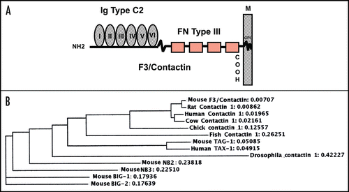
Figure 2 F3/Contactin expression profile. The F3/Contactin expression is shown in postnatal day 8 cerebellum (A), in newborn mice cerebral cortex, including the ventricular zone (VZ) (B) and cortical layers I–III (C). Expression in postnatal day 8 hippocampus is also reported (D). egl: external granular layer; igl: inner granular layer; PCl: Purkinje cells layer. I-II-III refer to the corresponding cortical layers. Scale bars: (A and D) = 200 µm; (B and C) = 40 µm.
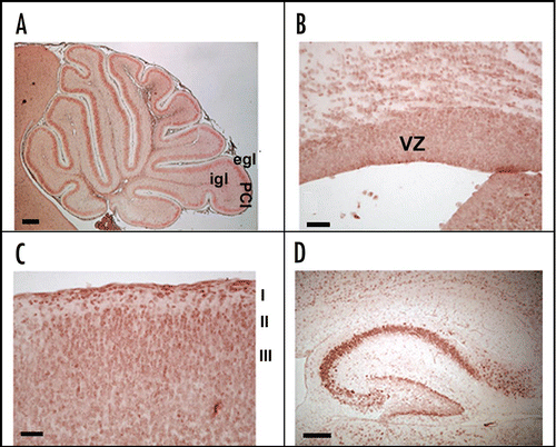
Figure 3 Activation of the F3/Contactin promoter in developing mouse cerebellum. (A) Organization of the 5′ flanking region of the mouse F3/Contactin gene. In (B) the 5′ flanking exons are shown, which undergo complex splicing events (shown in C), resulting in a high level of complexity of the F3/Contactin mRNA (reviewed in ref. Citation66). (D and E) Map of the F3/Contactin promoter/EGFP reporter construct (D), and its expression in developing cerebellum (E). Transgene expression recapitulates the endogenous gene with an earlier activation on migrating granule cells (P0, see also inset) and subsequent expression on Purkinje neurons (P8, see also inset). Egl, external granular layer; Igl, inner granular layer; PCl, Purkinje cells layer; WM, white matter. Scale bars: P0, P8 = 200 µm (insets 20 µm).
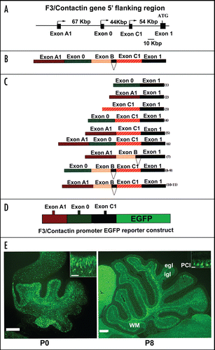
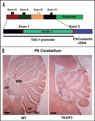
Figure 4 Changes in the cerebellar size arising from F3/Contactin developmental misexpression from the TAX-1 promoter. (A) Organization of the TAX-1 promoter/F3 cDNA construct. The F3/Contactin regulatory region, including the genomic sequences surrounding exons A1, 0, B and C1, has been replaced for by the promoter region from the human TAX-1 gene and used to drive the F3/Contactin cDNA in transgenic mice. (B) Changes in the cerebellar size arising from F3/Contactin developmental misexpression under control of the TAX-1 promoter, observed in postnatal day 6 mice. An F3/Contactin immunostaining is shown. egl, external granular layer; igl, inner granular layer; PCl, Purkinje cells layer; WM, white matter; CP, Choroid Plexus. Scale bar: 200 µm.