Figures & data
Figure 1 Contactins consist of six immunoglobulin-like domains and four fibronectin type III-like domains that are linked to the plasma membrane through a GPI-anchor.
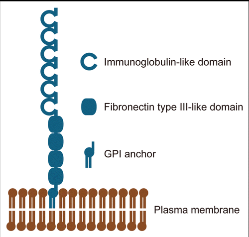
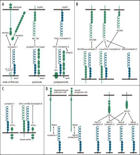
Figure 2 The interactions of contactins in a variety of tissues/cells/compartments. Contactins are drawn in blue, and other molecules are in green. (A) The molecular complexes in the myelinated nerves. (B) Interactions of contactins with molecules of the L1 family and NCAM. (C) Interactions of contactins with PTPα. (D) Interactions of contactins with Notch and molecules of the APP family.