Figures & data
Figure 1. Fluorescent staining of the f-actin cytoskeleton (green) and DNA (blue), showing cell spreading. (A) 6 h on MWCNTs. (B) 3rd day on MWCNTs. (C) 6 h on TCP. (B) 3rd day on TCP.
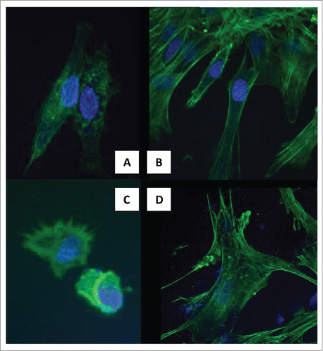
Figure 2. SEM images showing cytoplasmic processes and filopodia. (A–C) 3rd day on MWCNTs. (D) 3rd day on TCP.
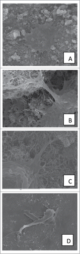
Figure 3. Quantitative real time RT-PCR gene expression analysis of vinculin gene.
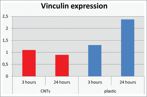
Figure 4. Lactate dehydrogenase (LDH) activity measured in cell supernatant after 24 h of culture on MWCTs and on TCP.
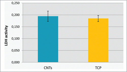
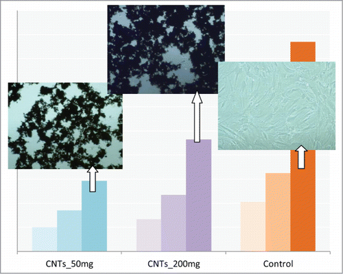
Figure 5. Total-protein of the hMSCs cells after 1, 3 and 7 d of culture on 2 different concentrations of MWCNTs. CNTs_50: 50 μg/mL, CNTs_200: 200 μg/mL and on TCP. Images of cultured cells on the substrates are also shown.