Figures & data
Figure 1 Transwell chemotaxis of SupT1 cells infected with viruses containing shRNA targeting genes encoding the indicated members of the Rab family or EGFP as a control. The number of cells migrating to the lower chamber was determined using CyQUANT as previously described.Citation1 Reductions in the chemokine dose response were significant using ANOVA (p < 0.01 for each of the knock-down cell lines).
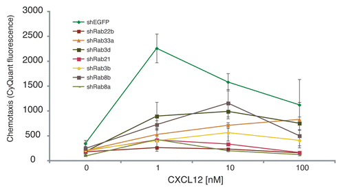
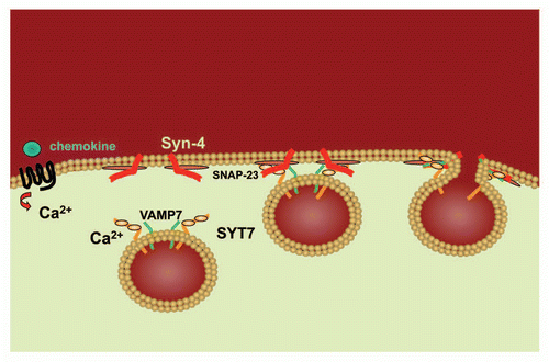
Figure 2 Model of SYT7 activity in chemotaxis. Following the binding of a chemoattractant (eg. chemokine) to its G protein-coupled seven transmembrane spanning receptor, calcium is released. Binding of calcium to Syt7 on the lysosome surface induces Vamp7 on the lysosome surface to interact with Syn-4 and SNAP-23 at the cell membrane thereby facilitating the fusion of the lysosome with the cell membrane.