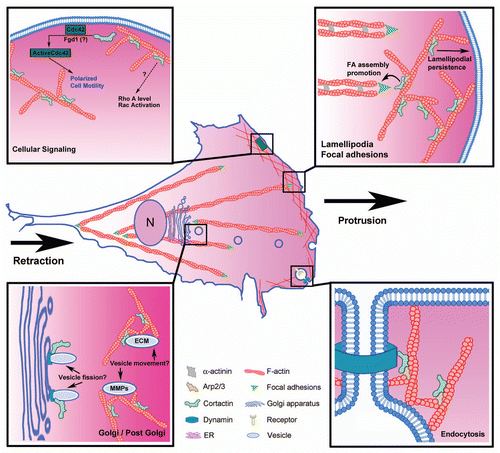
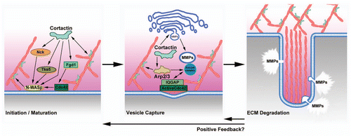
1,818
Views
133
CrossRef citations to date
0
Altmetric
Special Focus: Actin linked regulatory molecules

Cortactin

A multifunctional regulator of cellular invasiveness

, , &
Pages 187-198 | Received 02 Nov 2010, Accepted 11 Jan 2011, Published online: 01 Apr 2011

Figures & data

Figure 1 Regulation of cellular motility by branched actin and cortactin. Cell motility requires coordination of several processes, including protrusion of the leading edge lamellipodium, adhesion, contraction of actin bundles, and retraction of the rear of the cell. Depicted in the zoomed panels are mechanisms by which cortactin may regulate motility, including: promoting lamellipodial persistence, focal adhesion assembly, cellular signaling and secretion of autocrine factors

Figure 1 Regulation of cellular motility by branched actin and cortactin. Cell motility requires coordination of several processes, including protrusion of the leading edge lamellipodium, adhesion, contraction of actin bundles, and retraction of the rear of the cell. Depicted in the zoomed panels are mechanisms by which cortactin may regulate motility, including: promoting lamellipodial persistence, focal adhesion assembly, cellular signaling and secretion of autocrine factors

Figure 2 Cortactin domain structures. Schematic diagram of key cortactin domains and binding partners. The following abbreviations are used: NT A, N-terminal acidic domain and SH3, Src homology 3 domain. Proteins whose interaction with cortactin has been narrowed down to a particular domain are represented in the same color as the domain on cortactin. Interacting proteins shown in yellow bind the amino terminus of cortactin, which constitute the NT A + repeats domains. Amino acids that are essential for the interaction with key cortactin binding proteins, including W22 for interaction with Arp2/3 and W525 for interactions within the SH3 domain, are shown. The kinases known to phosphorylate cortactin are found above the respective sites they have been shown (or hypothesized) to phosphorylate.

Figure 2 Cortactin domain structures. Schematic diagram of key cortactin domains and binding partners. The following abbreviations are used: NT A, N-terminal acidic domain and SH3, Src homology 3 domain. Proteins whose interaction with cortactin has been narrowed down to a particular domain are represented in the same color as the domain on cortactin. Interacting proteins shown in yellow bind the amino terminus of cortactin, which constitute the NT A + repeats domains. Amino acids that are essential for the interaction with key cortactin binding proteins, including W22 for interaction with Arp2/3 and W525 for interactions within the SH3 domain, are shown. The kinases known to phosphorylate cortactin are found above the respective sites they have been shown (or hypothesized) to phosphorylate.

Figure 3 Model of cortactin function at invadopodia. Cortactin is thought to contribute to two major processes in invadopodia: (1) actin polymerization for initiation and/or maturation of invadopodia via activation of N-WASp via Nck, activation of cdc42 via Fgd1, and coactivation of Arp2/3 complex and (2) vesicular trafficking of matrix metalloproteinases to invadopodia via either regulation of post-Golgi trafficking or vesicle capture at invadopodia. Once ECM-degradation is established at invadopodia, they may become longer-lived due to positive feedback.

Figure 3 Model of cortactin function at invadopodia. Cortactin is thought to contribute to two major processes in invadopodia: (1) actin polymerization for initiation and/or maturation of invadopodia via activation of N-WASp via Nck, activation of cdc42 via Fgd1, and coactivation of Arp2/3 complex and (2) vesicular trafficking of matrix metalloproteinases to invadopodia via either regulation of post-Golgi trafficking or vesicle capture at invadopodia. Once ECM-degradation is established at invadopodia, they may become longer-lived due to positive feedback.

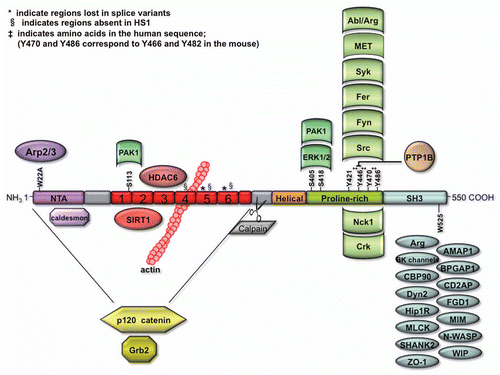
Table 1 Table of cortactin binding partners

Reprints and Corporate Permissions

Please note: Selecting permissions does not provide access to the full text of the article, please see our help page How do I view content?

To request a reprint or corporate permissions for this article, please click on the relevant link below:

Academic Permissions

Please note: Selecting permissions does not provide access to the full text of the article, please see our help page How do I view content?

Obtain permissions instantly via Rightslink by clicking on the button below:

If you are unable to obtain permissions via Rightslink, please complete and submit this Permissions form. For more information, please visit our Permissions help page.