Figures & data
Figure 1 Representative examples of immunohistochemically stained sections positive for EGFR (A and B), pAkt (C and D) and pERK (E and F) in tumor specimens ([A, C and E], x20 original magnification; [B, D and F] x40 original magnification). Scale bars represent 100 µm.
![Figure 1 Representative examples of immunohistochemically stained sections positive for EGFR (A and B), pAkt (C and D) and pERK (E and F) in tumor specimens ([A, C and E], x20 original magnification; [B, D and F] x40 original magnification). Scale bars represent 100 µm.](/cms/asset/9e540ea7-2dd1-4149-9b39-1eeaff2ab5c0/kcbt_a_10913877_f0001.gif)
Figure 2 Survival curves with Kaplan-Meier method of 102 ovarian cancer patients. Progression-free survival and overall survival of patients according to FIGO stage (A), EGFR gene mutation status (B), EGFR expression status (C) and pAkt expression status (D). p values were calculated using the log-rank test.
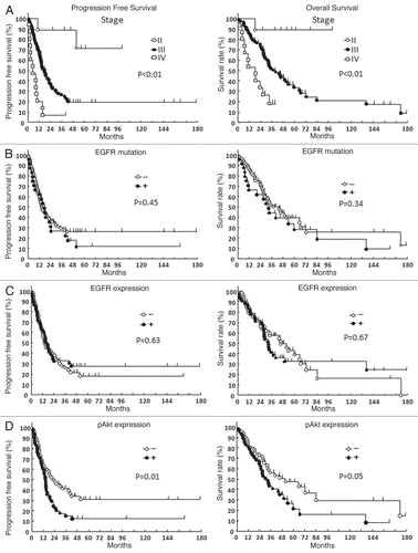
Table 1 Patient characteristics
Table 2 Type of EGFR gene mutations
Table 3 Correlation with clinicopathological features and molecular markers
Table 4 Multivariate analysis for survival rates